Category: Articles

  • The Devil Wears Science Journey

    The Devil Wears Science Journey

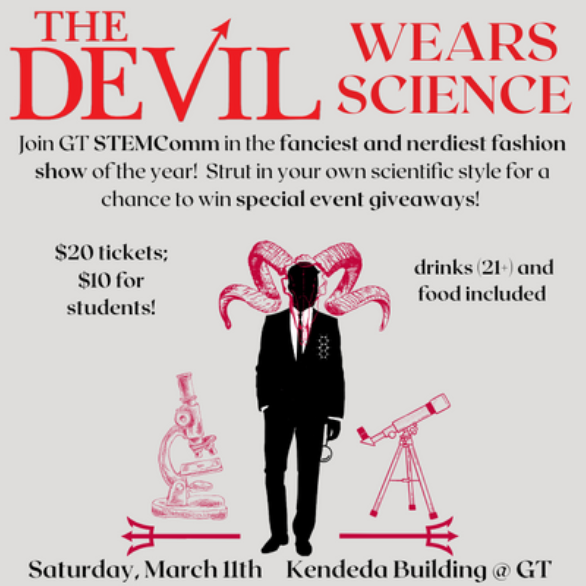
    Working behind the scenes of an event can be an eye-opening experience, especially when it’s a science show with a unique twist. That’s exactly what happened when I had the opportunity to be a part of the Atlanta Science Festival’s The Devil Wears Science fashion science show in March. Since it was my first time working as part of both a science show and fashion show, I was especially eager to be part of such a unique and innovative event. This immersive experience allowed me to gain valuable insights into the inner workings of a successful show, and I was amazed at the amount of hard work, creativity, and attention to detail that goes into putting on such an event.


    My group, Too Hot To Handle, focused on discussing and demonstrating the importance of heat stroke prevention through the use of protective clothing, specifically a beekeeping suit, during the event. My teammates were Claudia Ortega and Sachit Swaminathan and in the weeks leading up the show, we worked on reaching out to individuals in the beekeeping community to model and experts in the field of heat stroke prevention to present at the event, and also gathered information about the science behind our topic. 


    Prior to the event, I had very minimal knowledge about beekeeping suits, particularly as it relates to heat stroke prevention, so it was quite fascinating to do research and learn more about it. While there aren’t a large amount of scholarly resources on the topic, I tried to understand the different parts and materials of beekeeping suits and then did research on each fabric in relation to heat resistance. Moreover, ​​as I delved into the topic, I realized how much thought and consideration goes into selecting the right materials and design elements for gear such as beekeeping suits, especially since the consequences of inadequate protection could be severe. 


    There were some challenges my team and I faced during the planning of the event, particularly in regards to contacting people to discuss the science behind heat resistant gear. Initially, we planned for experts to discuss wearable heat sensor devices and then relate that to the design of heat resistant fabric in beekeeping suits, but we faced some difficulties in finding available experts to participate. After further attempts to contact other experts in the field, we decided to present the science portion of the presentation ourselves and focused on gathering research on the topic. 


    In addition to my work behind the scenes, I also had the opportunity to challenge myself and grow in my presentation skills. As the day of the festival approached, I decided to join Sachit in presenting the science portion of the event, which was a significant step out of my comfort zone. I conducted more research to develop a scientifically accurate script, and we kept making edits between the slideshow and script to allow the content to align. I was slightly behind at times due to preparing for a conference I had to attend later that week, but I was determined to gather all the necessary information and be confident in my speaking parts.


    On the day of the science fashion show, I was nervous as I arrived at the Kendeda building in my blazer clutching my freshly printed script.  Since there was some time to spare before the show began, I decided to use the opportunity to practice rehearsing for the presentation with my other teammates. This not only helped me to feel more confident and prepared, but it also allowed us to fine-tune the script and the visuals of our presentation, ensuring that everything was in sync and aligned with the overall vision of the show. As the presentation drew near, we gathered with our new models and went over the directions for modeling the beekeeping suits. Despite the initial setback of our previous model not being able to attend, we worked together and found a solution, and as we worked out other technical issues that arose, my confidence grew, and I became more and more assured in my part of the event.


    As our team took the stage and our presentation began, I couldn’t help but feel a sense of excitement mixed with a hint of nervousness. However, the more I spoke, the more my confidence grew and before I knew it, our part was over. The feeling of accomplishment and satisfaction was truly indescribable, and it was amazing to see all our hard work finally come to fruition. 


    But the night didn’t end there. As the other teams took the stage, the energy in the room continued to grow. I was captivated by the creativity and passion that each team brought to their presentations and it was incredible to see the range of scientific topics and the level of detail that went into each one. 


    Overall, I came away from this experience with a greater appreciation for the intersection of science and fashion, and the power of collaboration to bring together different perspectives and talents. It was an amazing opportunity to learn and grow and I look forward to participating in future events that combine my love of STEM and creativity.
     

  • 3 Free Applications for STEM Majors (That You’ll Actually Use)

    3 Free Applications for STEM Majors (That You’ll Actually Use)

    As STEM majors, if you look up helpful apps to help you while studying or writing papers, you’ll get a lot of listicles of apps that fall under a few categories:

    1. Costs money (or hidden costs)
    2. Does the same thing as a bunch of other apps. Most everyone already has a note-taking method, to do manager, or calendar that they already are comfortable using. Why use a new one?
    3. Way too technical and hard to understand

    Instead, I’ve compiled 3 apps that add value to your life in the following ways:

    1. Free
    2. Works as well/better than paid alternatives
    3. Takes less than 15 minutes to set up

    Let’s get it started!

    Anki

    How to learn anatomy with Anki: Flashcards, review | Kenhub
    An example of an Anki flashcard from kenhub.com

    I was team Quizlet since middle school, but the increased monetization has really driven me off the platform. This past year, I started using Anki, a free app that’s not only a good replacement, but an improvement.

    This app isn’t pretty–but if you look past its boring UI, you’ll find a gem of a tool. Here’s the highlights:

    • Basic UI + open-source software means it’s very customizable. With a few enabled settings, you can use it to study almost any kind of content: learning languages, doing physics practice problems, anatomy flashcards, and more. There are different mods you can add to your software to make it work better for you.
    • You can spread out studying over a longer period of time, so you retain info better!
      • Unlike Quizlet, Anki lets you set deadlines for cards depending on how well you know the content.
      • Naturally, you’ll end up studying the stuff you don’t know for much longer, saving you time!
    • So many stats: the app tells you which cards you’re better at, tracks the time you spend studying, and even notes which time of day you answer most accurately.

    Zotero

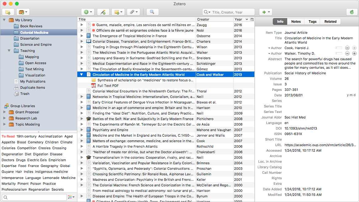
    Screenshot of the Zotero desktop application
    An example of a Zotero library from zotero.org

    No more keeping urls to your research sources in some random Word doc! Writing term papers and research essays has been one of my hardest struggles in college. It can be overwhelming to have to consume a bunch of research and analyze it in a meaningful way. Make it easier for yourself with Zotero, a “personal research assistant” that organizes sources for you. This tool is used by lots of people in academia, but not many undergrads know about it. Here’s what makes it great:

    • It’s easy to add sources! You can use the browser extension to add an entire source with just a click.
    • Once the source is added, all the information is downloaded: bibliographic information, tags associated with the work, a snapshot/pdf of the work
    • If there’s a pdf of the work, it’s saved locally to Zotero and you can take notes/highlight in the in-app pdf viewer!
    • Integrates with Google Docs and Word, where you can quickly add in text citations or a bibliography at the end of your writing

    ResearchRabbit

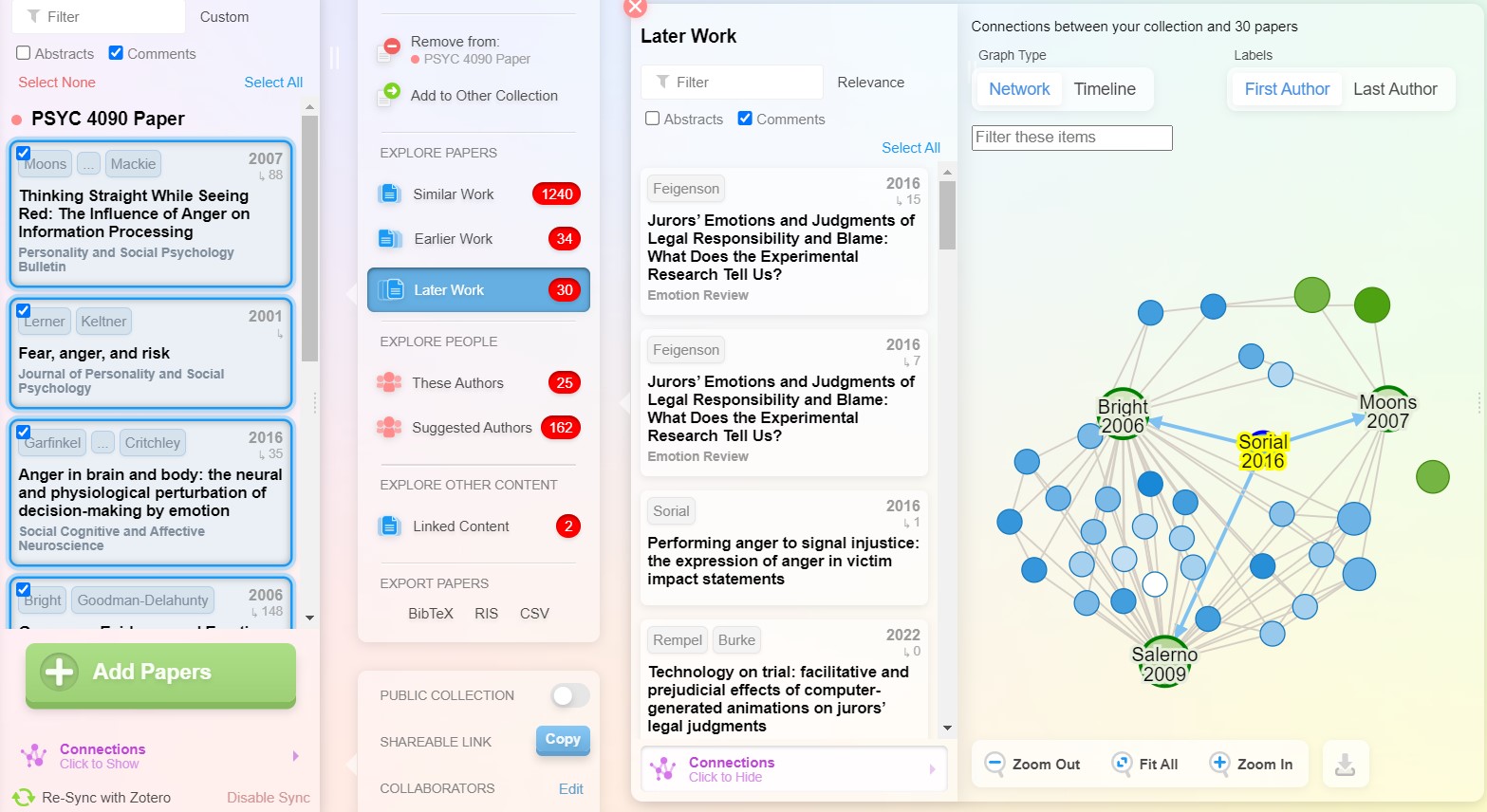
    A connection map that ResearchRabbit generated for one of my term papers.

    What does it mean to do a “thorough review of the literature”? That phrase can be daunting for undergrads. I don’t know about y’all, but when I’m usually starting a term paper, I don’t even know what I don’t know. Are these papers I’m using representative of the current research? This tool (researchrabbit.ai) gives you suggested papers, and it lets you see how popular your sources are in the field. Here’s my favorite features:

    • The coolest UI that makes it so easy to use
    • Visual maps help you “see” the impact of the sources you’re citing
    • The option to look at “similar work” and “related work” helps you find other sources, especially if you want to look at the current state of the literature


    These tools have been really helpful for me this year, and I hope they can help y’all too! Give these a go to help you work smarter, not harder. Good luck!

  • The Flower Power Behind Gene Silencing

    The Flower Power Behind Gene Silencing

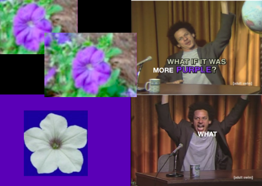
    Have you heard the story about some scientists trying to turn purple petunias more purple, and instead the petunias threw a tantrum and turned white?

    The theory I heard was that because the method to insert new genes into an organism usually involves using a modified virus, when the plant was “infected” with the extra purple, it’s “immune system” treated the gene like an attack and as a result just it shut down all together – and thus, white flowers.

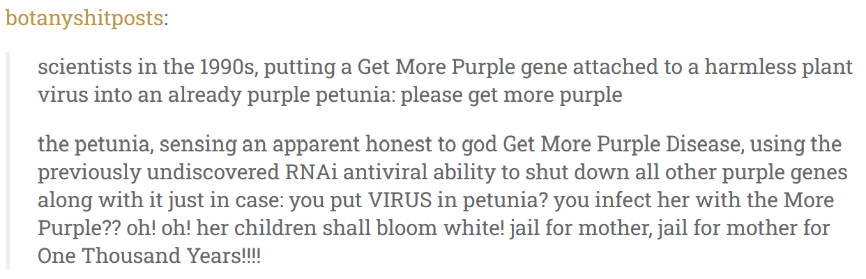
    Textpost that reads: "scientists in the 1990s, putting a Get More Purple gene attached to a harmless plant virus into an already purple petunia: please get more purple the petunia, sensing an apparent honest to god Get More Purple Disease, using the previously undiscovered RNAi antiviral ability to shut down all other purple genes along with it just in case: you put VIRUS in petunia? you infect her with the More Purple?? oh! oh! her children shall bloom white! jail for mother, jail for mother for One Thousand Years!!!!"

    IT TURNS OUT THOUGH, it’s a bit more complex than even that – and eventually, this little failed experiment led to the discovery of a major gene editing tool.

    So back in the 90’s, the scientist who tried to purple-fy the petunias (Richard Jorgensen) actually added the gene through transfection instead of transduction – essentially meaning they got the gene into the cell by non-viral means.

    HOWEVER, despite not actually being exposed to a virus, those petunias did respond like they were being attacked. This is because inserting foreign genetic material is exactly how viruses work, and in response, most organisms’ from plants to worms to humans, have developed protections against it.

    And this is especially true for RNA, since that’s what most viruses are made of – and what most cells need to defend against.

    A quick overview of RNA, for the unfamiliar. It’s a nucleic acid like DNA (“ribonucleic acid”), and it’s essentially DNA’s little brother. If DNA holds the blueprints for all the different proteins and things a cell needs to make, RNA is the way those instructions are translated and then messaged to the rest of the cell – AKA mRNA. This comes into play with viruses, because they function essentially like spam mail – they attack cells and add their own RNA instructions, which tricks and hijacks the host cell into making whatever the virus wants (usually more of itself). So cells have a defense to combat this.

    For one, mRNA on the job only has one strand (instead of 2 like DNA), so that it’s instructions can be read easily. However, nucleic acids come in pairs, so if another strand of RNA happens to come along that has the right matching pair, the two strands can get stuck together like legos – blocking the instructions and stopping the expression of that gene/protein. This is what researchers figured happened with the petunias, and several similar experiments with worms. However, another surprise occurred when researchers Andrew Fire and Craig Mello tried to do a control experiment where they inserted cells with a strand of mRNA that was supposed to have no effect – they still saw gene silencing! 

    Looking deeper into it, they found that the cause was actually double strands of RNA that accidentally formed before injection. Apparently these pre-paired RNA strands were even better at stopping gene expression than a single strand – which raised more questions as to why.

    And here’s where viral defense come in. Double strands of RNA (dsRNA) don’t really occur normally in cells, but they ARE common in certain viruses. So what these researchers discovered (and won a Nobel Prize for), was that plant and animal cells have a naturally occurring enzyme call Dicer that (as you might guess) finds and dices these dsRNA into little parts; and then it goes even further and takes those un-paired parts and uses them to hunt down and deactivate their partners – effectively erasing whatever virus, protein, or gene that was encoded in the targeted RNA. This is called the RNA interference pathway, or RNAi.

    Now it was known at this point that, at least in mammals, introducing long strands of dsRNA for potential gene therapies/medicines wasn’t really possible, since they often triggered a larger immune response. However, learning about this silencing method inside cells allowed scientists to determine that very small strands of dsRNA could be used to reduce and effectively erase a gene or protein. And these small interfering RNA (siRNA) have become key to creating groundbreaking new gene therapies, like treatments for debilitating genetic diseases, HIV, and potentially even cancer. 

    Quite a lot, to come from a seemingly failed flower experiment!


    https://en.wikipedia.org/wiki/Virus#cite_note-Collier9699-73

    https://www.independent.co.uk/news/science/how-an-experiment-to-change-the-colour-of-a-petunia-led-to-a-breakthrough-in-the-treatment-of-cancer-and-aids-5547175.html

    https://www.genengnews.com/insights/plucking-rnai-from-the-gene-silencing-nettle/

    https://faseb.onlinelibrary.wiley.com/doi/10.1096/fj.06-6014rev

  • Infectious Diseases, Immune System and Vaccines

    Infectious Diseases, Immune System and Vaccines

    Since ancient times the causes of severe infectious diseases in humans have been puzzling to people. Specifically, the recurrence of outbreaks, the severity of outcomes after infection ranging from permanent sequelae to premature death as well as the large impact in numbers in human populations have created the need to search for ways to learn about and prevent them. By continuous observations and through trial and error, civilizations were able to identify and distinguish the common features of certain infectious diseases and so to differentiate one from each other. Once fairly identified, people have been searching for effective and disease-specific ways to prevent them. It is in this search for prevention how vaccines appeared as tools against the nasty outcomes of infection. In this essay I will attempt a brief review of concepts related to how the immune system and vaccines work, and some key events in vaccine development.

    Infectious diseases with severe outcomes in humans are several and new diseases  continuously appearing. Yet, using the particular causes or the origins of infectious diseases, they can broadly be classified into four groups: bacterial, viral, parasitic and fungi. Among these causes, there are not yet vaccines against any parasite or fungus causing severe disease in humans. Among all those diseases caused by bacteria or viruses only a few of them have a vaccine to prevent humans from their burden. Even less, the available vaccines are only against a few strains, serotypes or serogroups of a given bacterium or virus. Therefore, the number of pathogens (bacteria and viruses) that could cause severe infectious diseases in humans is several times larger than the number of vaccines we have against them. 

    In general, bacterial and viral diseases are often grouped as pathogens causing diseases      

    Humans have been exposed to environments full of dangerous pathogens. These continuous exposure to pathogens caused outbreaks and diseases. The threat of infection requires response from human bodies and so the human body often reacts and fights the attacks of pathogens using an innate nonspecific immunity level, which is part of the immunity system. This is often called “primary response” which occurs following the first exposure to a foreign agent. If the response is quick and effective, the infection will be contained or eliminated. Sometimes, however, the response fails so infection would cause a disease.  This could happen when the human body’s immunity is low and/or when the pathogen has a virulence to overcome the response, rapidly multiplying itself inside the human body and causing great damage to cells and tissues.     

    When humans were or are able to survive and recover from certain infectious diseases, their bodies generate some specific level of immunity to reinfections. It is because the human bodies have an immune system which is essential for our survival and where some specific memory about previous infections is stored and keeps the defense abreast.  Specifically, that specific memory set in the immune system works as a first line of defense when potential reinfections with the same pathogens happen. Sometimes, such memory could wane in time and so make humans again susceptible to infection and disease; other times, exposure to the same pathogen would boost a decaying specific immunity and keep the protection against the attack of that pathogen high. Another important reason for humans becoming susceptible again is when the specific pathogen mutates and evolves, becoming unidentifiable and so making useless the previous specific memory of protection in our immune system.   

    Among all types of pathogens, the new or foreign ones of great concern are those that cause severe disease and often overwhelm the immune system by making their response slow and ineffective. Often are those that cause a heavy disease burden in the human population with plenty of sequelae or death. One way to prepare the immune system to such rapid and virulent attacks and to build a defense against them is with vaccines.  Although there are different types of vaccines even against the same pathogen, in general all of them work like sparring for box or combat fighters; namely, as a sparring does, a vaccine safely and effectively stimulates the immune system, trains its rapid response and prepares it to fight. Effective vaccines are often able to mimic some or all specific characteristics – also known as antigens– of the pathogen. To keep the training of the immune system safe, these antigens in the vaccine are often weakened or killed versions of the real pathogen. In average, during the first two weeks after vaccination, the vaccine is able to prime the immune system to respond fast and with force, should an infection with the particular pathogen in full strength happen in the future. Some refer to this process as programming of the immune system and to the rapid and specific response as “secondary response” or “vaccine-induced response.” An effective secondary response is often faster and with force in the production of pathogen-specific antibodies and in creating more cellular memory in the immune system for future attacks. Yet, vaccine’s protection also could wane with time or the specific pathogen could mutate and evolve. Similar to natural immunity that could also wane, vaccine immunity could also wane or become useless against new versions of the pathogen making human bodies susceptible again to infection.

    From the first vaccine developed in 1796 to the latest Covid19 vaccine, they are considered one of the greatest achievements of modern medicine. Throughout the last three centuries, several developments ranging from the advances in laboratory techniques, the discoveries in germ theory, the introduction of attenuated toxins, the cultivation of viruses, the evolution of cell culture to the introduction of recombinant DNA and the whole genome sequencing techniques have allowed the development of different vaccines and enjoy the progress and benefits we see today from them. In general, diseases that have caused the highest burden in morbidity and mortality in societies have received more attention and resources from those interested in developing vaccines. 

    Although vaccines have been an effective tool against infectious diseases, the development of each vaccine for a specific pathogen has often been a slow and research intensive process. Still, there were several attempts against some pathogens that did not end up with a safe and effective vaccine.  Vaccine development follows a rigorous scientific process focused primarily on safety and then on its effectiveness. All these processes should have a sound foundation on basic biology research, scientific discovery of vaccine candidates as viable proof of concept. This is the point when the application of the investigation of a new vaccine should be submitted. After approval, a three-phase clinical studies and trial begins.  

    Roughly summarized, Phase I is focused on vaccine safety conducting trials in small groups of both animals and humans; phase II is focused on vaccine effectiveness trials in an increased number of humans. Phase III is one of the most important ones. In this phase, thousands of individuals are recruited and the sample distribution should be representative of the population where the vaccine will be targeted. Individuals are then randomly assigned to often two groups  – this is referred to as random control trials or RCT.  RCTs often have a control group not exposed to the vaccine (control) and a trial group that will be exposed to the vaccine (trial group). Except for the exposure to the candidate vaccine, it is expected that both groups have very similar demographics and clinical characteristics to avoid confounding factors. Then, both groups are followed and clinical and lab data collected. By comparing the rates of disease cases in the exposed group with those of the control group is how the vaccine safety and effectiveness are determined. If successfully safe and effective, the vaccine data is then submitted for analysis by independent institutions and then the vaccine is licensed and recommended.     

    In the recent past, the research and development of new vaccines used to take about 15 to 20 years. However, thanks to more than three decades of advances in DNA sequencing and the stable production of proteins in human cells exposed to messenger RNA (mRNA), the research and development time has been reduced substantially and made possible the development of Covid19 vaccines in less than a year. Had the Covid19 pandemic occurred 30 years ago, we would have been in worse shape to control or prevent their consequences.  

    Overall, human’s immune systems could rely on more effective and safe vaccines to prime their response against pathogens’ attacks. Yet, the burden of infectious diseases could only be partially reduced by vaccines. As mentioned above, among all those diseases caused by bacteria or viruses only a few of them have a vaccine to prevent humans from their burden and for those with a vaccine, the protection is often partial and may wane in time.   


    *Note: All the ideas developed in this essay were based on the references listed below.  

    References:

    1. “A Brief History of Vaccination.” World Health Organization, World Health Organization, https://www.who.int/news-room/spotlight/history-of-vaccination/a-brief-history-of-vaccination. 
    2. Coon, Lisa. “The History of Vaccines and How They’re Developed.” OSF HealthCare Blog, 11 Feb. 2022, https://www.osfhealthcare.org/blog/the-history-of-vaccines-and-how-theyre-developed/. 
    3. Diseases.” History of Vaccines RSS, https://historyofvaccines.org/diseases.
    4. Dolgin, Elie. “The Tangled History of Mrna Vaccines.” Nature News, Nature Publishing Group, 14 Sept. 2021, https://www.nature.com/articles/d41586-021-02483-w. 
    5. Offit, Paul. “Developments by Year.” Children’s Hospital of Philadelphia, The Children’s Hospital of Philadelphia, 20 Nov. 2014, https://www.chop.edu/centers-programs/vaccine-education-center/vaccine-history/developments-by-year. 
    6. Saleh A, Qamar S, Tekin A, Singh R, Kashyap R. Vaccine Development Throughout History. Cureus. 2021 Jul 26;13(7):e16635. doi: 10.7759/cureus.16635. PMID: 34462676; PMCID: PMC8386248. https://www.ncbi.nlm.nih.gov/pmc/articles/PMC8386248/#:~:text=The%20first%20trials%20to%20develop,the%20United%20States%20in%201945. 
    7.  “U.S. Vaccine Safety – Overview, History, and How It Works.” Centers for Disease Control and Prevention, Centers for Disease Control and Prevention, 9 Sept. 2020, https://www.cdc.gov/vaccinesafety/ensuringsafety/history/index.html. 
    8. “Vaccine Development, Testing, and Regulation.” History of Vaccines RSS, https://historyofvaccines.org/vaccines-101/how-are-vaccines-made/vaccine-development-testing-and-regulation.
  • What’s It Like Working in a Mouse Lab?

    What’s It Like Working in a Mouse Lab?

    This semester, I started doing animal work in a research lab that uses mice as model organisms. Every time I’ve explained my training to other people, I usually get a shocked response. And I know this isn’t a common thing people do, so I wanted to give y’all a look into what it’s like working in an animal lab.

    The lab I work in does research using a technique called optogenetics. Opto- means light, and genetics refers to the DNA that is changed. This might sound straight out of a sci-fi novel, but by infecting a certain brain area of healthy mice with a special virus (this doesn’t make the mice sick, by the way), the virus can insert a list of instructions written in DNA into the mouse’s own DNA. These instructions tell the cells to start producing light-sensitive proteins in their membranes.

    Now, this has a very important application in neuroscience. Neurons, the unit cell of the brain, fire tiny electrical signals called action potentials. If a neuron has light sensitive proteins that respond to blue light (channelrhodopsin), it fires more often. If it has light sensitive proteins that respond to yellow light (halorhodopsin), it fires less often. Using optogenetics, you can directly measure how activation of a brain area leads to behavior in real time.

    So how does that factor into what I do? So far, I’ve worked on an often-overlooked but important step: collecting samples. After you inject the virus into the brain, there’s no way to check in the live animal that you did it right. You might have put the virus in the wrong place, or the virus never got its instructions to the cells, or the equipment might be broken. The only way to know for sure that your procedure worked is to take samples of the brain, and look at them under the microscope.

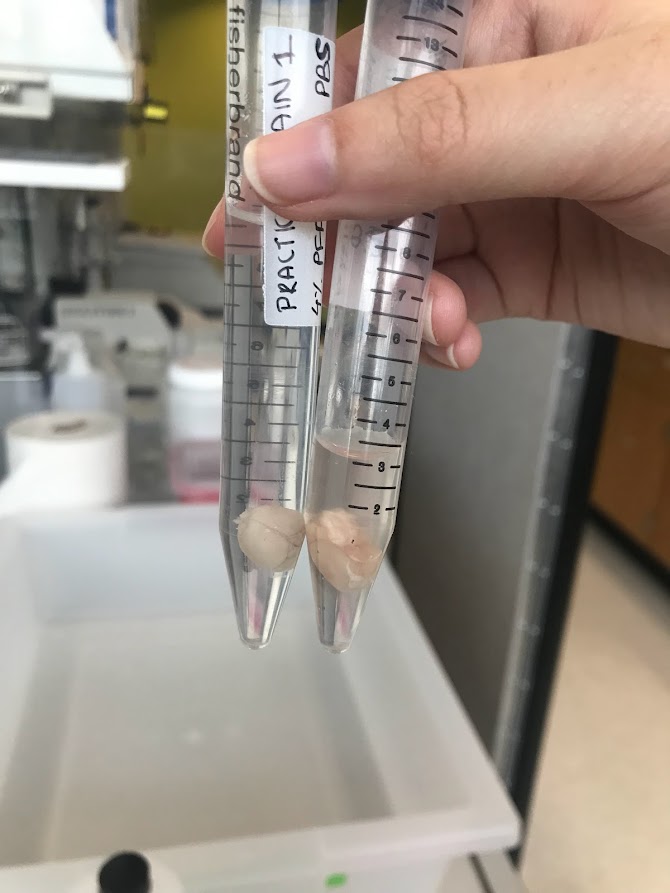
    To do that, we first need to collect the brains. Through a process called perfusion, the brain gets fixed in paraformaldehyde (PFA), a safer version of the formaldehyde you might remember from dissections in biology class. During drop fixing, where the brain is severed from the spinal cord and removed from the head, we store it in a test tube with more PFA.

    Two mouse brains in a solution of PFA, each in its own test tube.

    After 48 hours, the brain is ready to be sliced. There’s a lot of ways to take brain slices, but we use a vibratome. First, we locate the area of the brain that we’re trying to reach. This is where a mouse atlas comes in handy. Using a small, vibrating blade, we make slices of 50 microns–or around the width of a human hair! These slices are then stained with dyes like DAPI (which makes neurons look blue)  and placed on microscope slides.

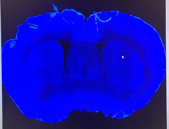
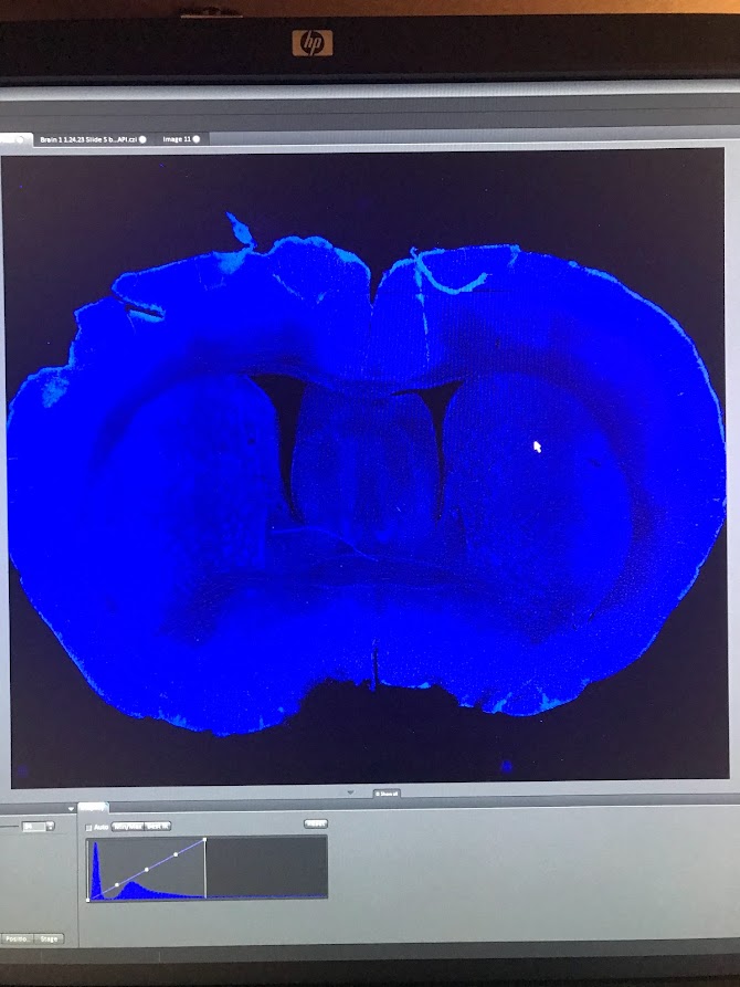
    Finally, after all this work, we can see the fruits of our labor. In our lab, we use a confocal microscope because this type of microscope can show us thin layers of cells within thick layers of tissue. Using different colors of light, we can also check how well the light sensitive proteins were able to grow in the area of the brain we’re interested in. In the picture below, you can see the first picture that I took myself from a brain I perfused and sliced. There’s no injection in this brain, so it’s as close to a “natural” brain slice that you can get. The blue color is from the DAPI dye, which indicates the location of DNA in a nucleus. So, each tiny blue dot is a neuron.

    A microscope image of the coronal slice of a mouse brain, with thousands of tiny blue dots representing individual neurons.

    It was hard to describe the first time I saw this image. Yeah, there were some nicks and tears in the tissue (my hands were shaky during the perfusion), but this was something that I had wholly produced myself. And now, in front of me, was a snapshot of all that this mouse was. 

    See that little butterfly shaped area in the middle of the brain, right above the hole in the middle? That’s the hippocampus, where every memory is scrawled in your brain through a network of neurons. What mousy memories were kept in there? Looking at this picture, I can almost imagine what it would have been like in there when the mouse was alive. The holes in between and to the side of the “wings” are ventricles, gaps in the tissue where cerebrospinal fluid carrying nutrients and hormones was kept. I can almost imagine waves of it washing over the hippocampus, keeping this great curled machine firing, shooting sparks of electricity in patterns that say I am alive. I have lived. Here is what I have seen, heard, smelled, touched, tasted, hated, loved. 

    To be honest, this work can be kind of sad. After all, all life is precious. Sometimes, it can feel almost gruesome. To perfuse a mouse, you stick a needle of fixative into its beating heart, so it evenly diffuses and preserves the entire body. The night after I read this protocol, which had a video of this process in high definition, I felt almost too nervous to sleep. When I closed my eyes, I just saw that mouse heart beating out of its chest, beating, beating, beating…

    The next morning, I admitted to my principal investigator that I wasn’t sure about trying out the perfusion that day. “Everything else, I’m okay with, but the way you can see the heart pumping freaks me out. I still want to try it, but…”

    He nodded. “You never really feel comfortable with this part of the work.”

    After a moment, he added, more seriously, “and that’s a good thing.”

    Testing on animals is a controversial subject, and I can’t claim to have the definitive opinion. But, I think my experiences give me a special point of view. In the animal labs at this institute, the animals are well cared for. Until the last day they’re with us, we treat them with respect, care, and even love (what else do you call grown adults cooing at a 2 inch long mouse washing its tiny little face with its tiny little paws?). There’s laws, regulations, committees to ensure that animals are treated ethically. They’re given pain medications after surgeries, and during procedures (surgery and perfusions) they’re put under anesthesia. Once, our lab manager stayed in the surgery room with the mouse she was operating on until night fell to make sure she was there to care for him when he woke up. 

    And yet, we have to sacrifice these animals in the end, and we have complicated feelings about it. I think that’s good and necessary. If we feel sad at the animal’s death, we treat it well when it’s alive. If we feel a bit of horror at watching its life painlessly bleed away in the dissection pan, it means we placed a high value on that life in the first place. We try to perfect our surgical skills, write better protocols, and design better experiments to minimize mistakes and the number of mice who have to undergo this process. I would much prefer that than to become cold and callous. 

    Because the truth is, we don’t have a better process to study the brain in vivo. Treatments for neurodegenerative disorders, like Parkinson’s and Huntington’s (two diseases targeting the brain area our lab studies), mental illnesses, and mental disorders lag far behind those for many other parts of the body. And the simple reason is that we often can’t get into the nervous system to study it without causing permanent damage. Until better options arise, this is the best way to perform this life-saving research.

    Whew. Things got really serious there. But working with something as big as life and death all the time makes you think about big questions. For me, ultimately this type of research is justifiable: this is the only way to validate that the research being conducted is working properly. It’s the only way to make sure that all the weird situations we put mice in weren’t done in vain. And getting to do every step of this process, going from a live mouse to a bright image on a screen–that’s something really special. Through that intricate picture, you can almost see something persisting, living on…helping us unlock the secrets of the brain. There is something to be said, here, about loving something by studying it: giving it all your attention while taking notes, appreciating the simple symmetry of the brain slice, using the gentlest touch to mount delicate tissue onto slides, admiring the natural structural variations that nature gifts every body and every brain.

    Finally, I want you to think about your daily life, and the laboratory animals that made it possible. How many medications did you take today? What about cosmetics? Vaccines? Animal testing was where many of them began. One funny thing I’ve noticed recently is that researchers who work with “animals” tend to be the most adamant about using the term “humans and non-human animals”. Other species of animals are intertwined with us in inextricable ways. And there’s really no way to really convey my gratitude to a species that can not understand it. 

    I guess this article is my attempt to try.

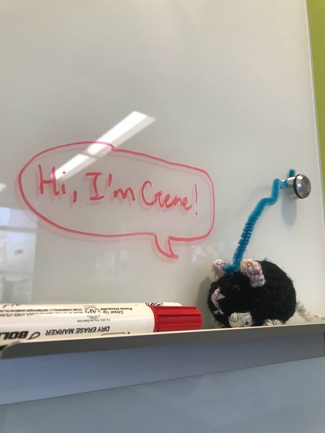
    A crochet mouse sits on the marker holder of a white board. On the white board, a text bubble reads, "Hi, I'm Gene!"

  • The Brachistochrone and Snell’s Law: A Surprising Connection

    The Brachistochrone and Snell’s Law: A Surprising Connection

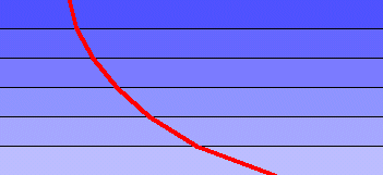
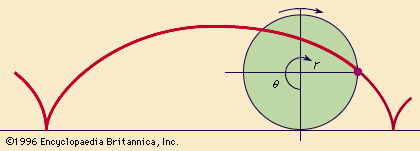
    Brachistochrone – A word originating from Greece that means “shortest time.” The brachistochrone problem is a well known problem in the world of physics that involves finding a curve such that, assuming there is no friction, a ball traveling along this curve would reach its destination in the shortest possible time. Initially discovered by Johann Bernoulli, a famous Swiss mathematician, the brachistochrone curve has a variety of interesting properties, such as the fact that it is a cycloid, the path mapped out by a point on a circle as it rolls on a flat surface. Additionally, a subsection of the curve also acts as a tautochrone curve, a curve that causes a ball to reach its destination in the same amount of time regardless of where it begins its journey. However, perhaps the most interesting fact about the brachistochrone is that it was initially solved by Bernoulli using Snell’s law, a law that dictates the path traveled by light when moving between different mediums.

    A cycloid is mapped out in red. When inverted, this curve matches the shape of the brachistochrone.

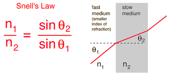
    Initially, it may not be readily obvious why Snell’s law would approximate a brachistochrone curve, but with a bit of physical intuition, it makes perfect sense. First, the reason why Snell’s law works must first be understood. Snell’s law states that the ratio of the sines of the angles formed by the refraction of light when passing through a medium is equal to the ratio of the speed within the two media.

    The index of refraction dictates how much light “slows down” in a medium.

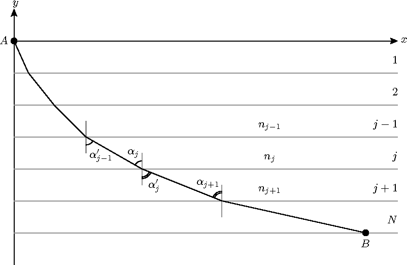
    With a bit of calculus not shown here (perhaps in a future article), it can be shown that this equation comes as a consequence of minimizing the time taken for light to travel from one point to another between the two media. This is because light naturally always takes the path of shortest time, and this is what connects Snell’s law to the brachistochrone. When a ball is dropped, it is constantly changing velocity due to gravity, so you can envision a ball as traveling through a continuous set of layers of different velocities. If the ball wanted to travel the path of shortest time, it would follow Snell’s law at every point in its path, so the brachistochrone curve should have the property of having the ratio of instantaneous change in the velocity of the ball match the ratio of the angles formed by a vertical line at that point of the curve.

    Here, the line travels through several “layers” with different speeds due to gravity and diffracts according to Snell’s law each time.

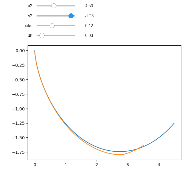
    Of course, it’s one thing to be told that Snell’s law maps out the brachistochrone, but it’s not as satisfying as seeing it for yourself, which is why I coded a simulation in Python to try and match up a curve constructed from Snell’s law with a brachistochrone curve.

    The top two sliders change the initial conditions of the actual brachistochrone in blue, while the bottom two sliders change the initial angle and the number of layers for the Snell’s law approximation in orange.

    For more information on the brachistochrone curve, check out Vsauce or 3Blue1Brown’s videos on the topic below:


  • Researching the Anterior Cruciate Ligament

    Researching the Anterior Cruciate Ligament

    This semester I took a course called Locomotion Neuromechanics. Like me, you might think that it would be a course where you would learn more about the movement within one’s body, different things that can affect mobility, etc. However, after attending the first class, I realized it was focused on the student’s own interest in a topic relating to Locomotion Neuromechanics. I found this method interesting because it allows the student to either dive deeper into their own research or explore other topics, related to locomotion, that they wouldn’t be able to in another class.

    After the first round of presentations, the class had a broad range of topics that would be focused on like robotics, animal/insect locomotion, neurodegenerative diseases that impact mobility, flying snakes, spinal cord injuries, etc. At first, I had no idea on what topic I would be focusing on. I initially presented on something related to spinal cord injuries, but instead of focusing on humans, I was looking at animals like cats and mice. This changed after I was researching stuff that I would enjoy more. I then focused on the anterior cruciate ligament or commonly known as the ACL. This topic can go any way from what is the ACL, how it works, ACL injuries, etc.

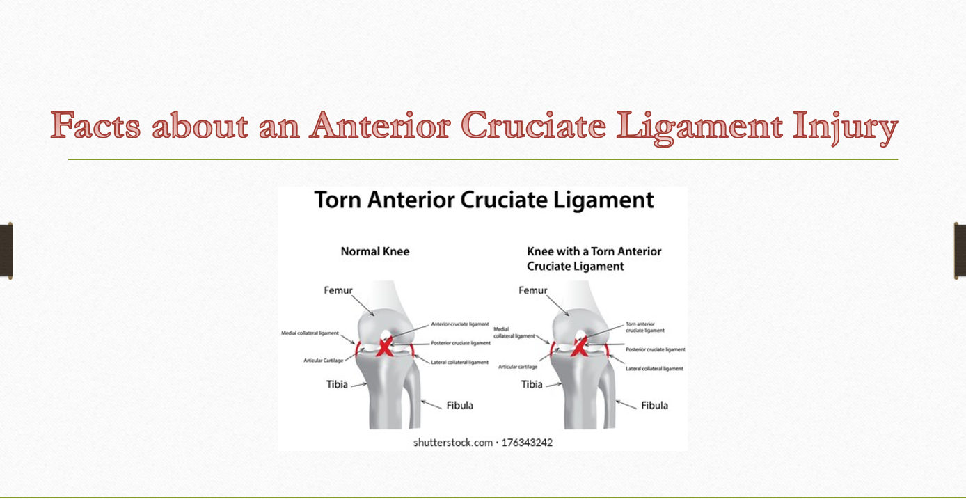
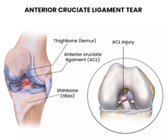
    Before I start, you might be wondering what the ACL is. The ACL is a ligament that connects the femur to the tibia. The femur and tibia in layman’s terms are the thigh bone and shinbone, respectively, found in your leg. This ligament is important for knee and joint stability. You might wonder why your knee doesn’t move forward when you walk, jump, play sports, etc. The ACL is in charge of preventing the movement of the knee to go from the front to the back or side to side. It is most commonly known to have ACL injuries while playing sports such as soccer, football, basketball, gymnastics, etc.

    Below are some of the interesting research facts that I found on my literature search about the ACL:

    • The two most common ACL reconstruction surgeries: bone patellar tendon bone graft and the hamstring graft
    • Hamstring strength recovered at a faster rate than quadricep strength (3)
    • Anterior knee pain influences the recovery of muscle performance of a patient after ACL reconstruction (3)
    • Younger patients had a higher rate of subsequent ACL injury rate, distinctly that young males had higher rate of rupture than females (6)
    • Researchers are trying to focus on rehabilitation programs that focus on the different forces that might heal an ACL (6)
    • Depending on some factors like the type of graft used, how the graft works post-operatively and the different fixation techniques (6)
    • In addition to rehabilitation programs, literature states that they are examining pre and post operative rehabilitation, neuromuscular training, electrostimulation and other protocols that an individual can go through after ACL reconstruction. (5)
    • A long term consequence of ACL reconstruction is osteoarthritis which is known to be a disease of the joints. Factors that can influence the outcome consists of sex, age, family history and muscle strength (4)

    These were just a couple of interesting literature facts that my research came across focusing on ACL before and after reconstruction surgeries.
    I am working on the final project for this class so if anyone is interested in other facts about the ACL or interested to learn more, let me know.


    References:

    1. Cavanaugh JT, Powers M. ACL Rehabilitation Progression: Where Are We Now? Curr Rev Musculoskelet Med. 2017 Sep;10(3):289-296. doi: 10.1007/s12178-017-9426-3. PMID: 28791612; PMCID: PMC5577427.   https://pubmed.ncbi.nlm.nih.gov/28791612/
    2. Freeman. “ACL Injuries.” NJ’s Top Orthopedic Spine &; Pain Management Center, 7 Mar. 2023, https://redefinehealthcare.com/acl-injuries/.
    3. Kobayashi A, Higuchi H, Terauchi M, Kobayashi F, Kimura M, Takagishi K. Muscle performance after anterior cruciate ligament reconstruction. Int Orthop. 2004 Feb;28(1):48-51. doi: 10.1007/s00264-003-0502-5. Epub 2003 Aug 26. PMID: 12942198; PMCID: PMC3466568. https://doi.org/10.1007/s00264-003-0502-5
    4. Lohmander, L. S., Englund, P. M., Dahl, L. L., & Roos, E. M. (2007). The long-term consequence of anterior cruciate ligament and meniscus injuries: osteoarthritis. The American journal of sports medicine, 35(10), 1756–1769. https://doi.org/10.1177/0363546507307396 
    5. Perry A, DeFroda S, Leporace G, Metsavaht L, Coxe CR, Bierman AM, Chahla J. ACL Rehabilitation: How Can We Lessen Injury Rates? Operative Techniques in Sports Medicine, 30,1, 2022. ISSN 1060-1872, https://doi.org/10.1016/j.otsm.2022.150892. https://www.sciencedirect.com/science/article/pii/S1060187222000041
    6. Webster KE, Feller JA. Exploring the High Reinjury Rate in Younger Patients Undergoing Anterior Cruciate Ligament Reconstruction. Am J Sports Med. 2016 Nov;44(11):2827-2832. doi: 10.1177/0363546516651845. Epub 2016 Jul 7. PMID: 27390346.  https://pubmed.ncbi.nlm.nih.gov/27390346/
    7. https://www.shutterstock.com/image-illustration/torn-anterior-cruciate-ligament-176343242