Category: Articles

  • What’s in a ‘species’? Discussing the Colossal Dire Wolf

    What’s in a ‘species’? Discussing the Colossal Dire Wolf

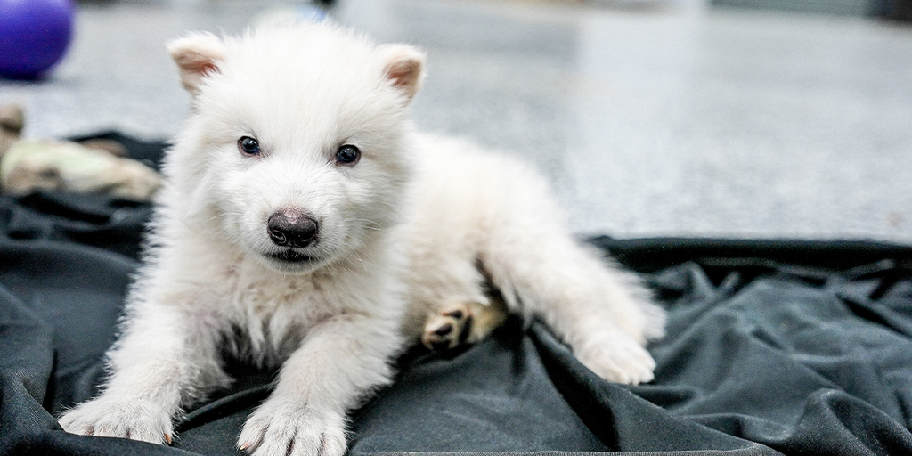
    April 2025. Did Colossal bring back the dire wolf?

    Short answer, not really.

    The long answer is a bit more complicated and goes into what ‘species’ actually means.

    Earlier in April, the biotech company Colossal put out a press release announcing the first de-extinction. They’ve genetically engineered a dire wolf out of extinction—and now, for the first time in 10,000 years, they’re back in their natural habitat (or so Colossal says).

    De-extinction is the process of bringing back extinct species, or modern-day analogs to extinct species. This can be done through cloning DNA from preserved specimens or by gene editing their closest related surviving species to make them resemble their extinct kin. While this used to be an unlikely science-fiction dream, the development of CRISPR-Cas9 and other gene editing techniques has made it a possibility.

    Colossal says their team used DNA sourced from a 13,000 year old tooth & 72,000 year old skull to analyze the dire wolf’s genome. They used this as the basis for their edits, which they carried out on three gray wolf embryos.

    What is a species?

    If you’ve taken a biology class, you’ve probably heard the term “species” tossed around before. The conventional definition of species is a group of organisms in which two individuals can reproduce & create fertile offspring. It’s generally a good rule of thumb definition, but there’s a lot of edge cases where the topic becomes muddy.

    What happens when you look at critters that only reproduce asexually, like bacteria? What about cases where two species hybridize, and remain fertile?

    These outlier cases have led some scientists to propose alternate definitions for species. Colossal’s Chief Science Officer (CSO) Beth Shapiro said in an interview with ABC:

    “I think that the best definition of a species is if it looks like that species, if it is acting like that species, if it’s filling the role of that species then you’ve done it.”

    This definition of species depends on its phenotype, behavior, and niche. A phenotype is a species’ physical expression of its genes. For instance, your eye color is a phenotype and is the observed effect of the genes behind your eye color. Shapiro said that their analysis of ancient dire wolf DNA suggested that they had light fur, which is why Colossal edited their gray wolf skins to be an eye-catching white. But instead of editing the pigment genes actually responsible for fur color, the team instead disabled two genes whose absence in dogs & other canids is known to cause a white coat. Colossal avoided the actual edits because they were linked to increased risks of deafness, blindness, and albinism—traits that the team didn’t want to take chances on.

    What about behavior? Does the dire wolf behave like a real dire wolf? Well, they’ve been extinct for thousands of years, and we lack any actual evidence as to their behavior, so we can’t actually test that point.

    What about their niche? A niche refers to the ecological role a species carries in its environment. For example, owls occupy the niche of eating small animals such as mice in woodland areas. The dire wolf was a carnivore that likely scavenged & hunted large megafauna. It was a species adapted for life in Pleistocene, and part of why it went extinct is thought to have been the extinction of its typical prey. If a dire wolf was to be de-extincted & rewilded today, it would be in unfamiliar territory—there are no more megaherbivores. Additionally, the three wolves Colossal has bred are living in a fenced-off lot and aren’t actively contributing to their environment.

    Following Shapiro’s definition of a species, Colossal’s dire wolf checks the phenotype box but doesn’t meet the behavior/niche ones. Is that enough to consider it a species?

    At the end of the day, the idea of what a species is is complicated. Ambiguity & argument around definitions like this can hold back science. This linguistic debate is no trifling matter—it can impact real world legislation and policy! For example, as soon as the news of Colossal’s ‘de-extinction’ broke, the Secretary of the Interior Doug Burgum tweeted out a statement suggesting that now that de-extinction technology is viable, the Endangered Species List is on the chopping block. De-extinction might be used to devalue the importance of conservation, instead shifting the mindset of preservation to one of “we can just bring them back!”

    If you look at the news, you’ll notice a lot of the reporting around the Dire Wolf story focuses on the miracle of “listening to the howls of a dire wolf pup brought back from extinction for 10,000 years.” By focusing on the info that brings the best headline, science can sometimes be shifted out of focus.

    Whenever new technologies come out, it’s important to approach them with a critical mind. What do you think defines a species? Do you think Colossal has revived the dire wolf?

    Works Cited

    https://www.technologyreview.com/2025/04/08/1114371/game-of-clones-colossals-new-wolves-are-cute-but-are-they-dire/

    https://abcnews.go.com/US/dire-wolf-revived-biotech-companys-de-extinction-process/story?id=120558562

    https://evolution.berkeley.edu/evolution-101/speciation/defining-a-species/

    https://x.com/SecretaryBurgum/status/1909345951069651032

    Picture credit Colossal

  • Friendly Fires

    Friendly Fires

    As climate change gets worse, more and more reports of disastrous forest fires have been hitting the news. But not all fires in the woods are bad—some of them can actually be instrumental in keeping an ecosystem healthy!

    Fire ecology is the study of how wildfire interacts with the environment. The field acknowledges the idea that fire is a natural part of the ecosystem and promotes change. It can clear away leaf litter and plants to provide nutrients and space for new growth.

    One of the best and most well documented examples of a fire-dependent species is the longleaf pine. Once covering over 92 million acres across the US, heavy logging had reduced it to just 3% of its natural range by 1992. The pine grows fast, thin, and tall, and has evolved to rely on fire to give it a start.

    longleaf pine | Roads End Naturalist
    Young longleaf pines burned in a fire

    In order for its seeds to germinate, the tree relies on starting on a forest floor free of any leaf litter. After the seed germinates, it grows a into a dense cluster of pine needles, low to the forest floor. These needles help provide fire resistance to the plant, protecting its bud from any fires. Its bud grows rapidly, allowing it to quickly grow above typical flame heights.

    Fire enables its growth by clearing the forest of competition from hardwoods and other pines. It maintains conditions best suited for longleaf growth and recycles nutrients into the soil. Fire also allows helps clear out insects and pests that can impede plant growth.

    Longleaf pines aren’t fireproof—they can still burn in its early stages of growth. But they are incredibly fire resilient. Mature longleaf pines have thick, plated sheets of bark that protect the trunk from fire. The canopy is usually high above the forest floor, enabling it to avoid fire altogether.

    Nowadays, the US Forest Service encourages and carries out controlled burns to ensure our remaining longleaf forests remain healthy. But this wasn’t always the case. When America was being colonized, early settlers and loggers didn’t know about the role of fire in longleaf ecosystems, and ignored indigenous knowledge that touted its benefits. Fire prevention became the forestry ethic to live by, because it felt paradoxic to burn the forest on purpose to keep it healthy. But as soon as they began preventing fires, they realized that naturally occurring forest fires became more disastrous.

    Because there weren’t frequent, little fires clearing out the forest, the fuel load, or flammable content, of the forest floor would keep growing. When it hit critical mass and finally caught fire, the fires would be massive and even harder to control. A lack of fires also led to a lower biodiversity. The fire habits of longleaf ecosystems leads to one of the highest species-per-acre densities—it’s been known to reach 40 species in a single square meter! Red-cockaded woodpeckers, indigo snakes, Bachman’s sparrow, flatwoods salamanders, sandhill lilies, and Sherman’s fox squirrel are just a few species that rely on longleaf ecosystems, which wouldn’t be possible without fire.

    To see a really cool visual of a longleaf pine stand’s growth over a year, check out this story map created by the Longleaf Alliance.

    Works Cited

    https://www.fireinthepines.org/learn/the-longleaf-ecosystem/

    https://longleafalliance.org/what-is-longleaf/restoration-management/prescribed-fire/

    https://www.nclongleaf.org/burningLongleaf.html

    https://www.nature.org/en-us/about-us/where-we-work/united-states/north-carolina/stories-in-north-carolina/north-carolina-role-of-fire-in-longleaf-pine-forests/

    https://bittersoutherner.com/feature/2024/burning-the-forest-for-the-trees-longleaf-pine-controlled-burn-janisse-ray

    https://www.nps.gov/articles/learning-about-fire-ecology-basics.htm

    https://longleafalliance.org/what-is-longleaf/the-ecosystem/built-by-fire/

  • Lie Detector Pseudoscience: Why Polygraphs Fail

    Lie Detector Pseudoscience: Why Polygraphs Fail

    How often have you heard a mention of a machine called a “lie detector?” According to pop culture, members of law enforcement organizations, and even the U.S. government, a polygraph or lie detector is a machine that records certain vital functions of a person suspected of lying. The person is asked a series of questions while the polygraph reads their physiological responses, and the resulting readings are supposedly highly accurate evidence as to whether that person was lying or telling the truth.

    Over the decades, many people have protested the use of the polygraph in the justice system, as a tool for detecting disloyalty in intelligence agencies, or as a condition of employment. Some arguments have been based mainly on philosophical objections to the concept of a lie detector: Preemptive polygraph tests contradict the principle that people are innocent until proven guilty in a fair justice system, or they violate a person’s constitutional right to be protected from self-incrimination, or they prevent them from “remaining silent” regarding criminal charges. These arguments have merit, and they are undoubtedly made out of genuine concern for human rights. However, there is a much more practical, relevant, and immediate reason to protest the use of polygraphs as lie detectors: They don’t work.

    The basic premise that is used to promote the accuracy of polygraphs is that all humans – regardless of age, sex, ethnicity, health, personality, and all other factors – display some uniform,  measurable physiological reaction when saying something they know to be false which is clearly different from their response to stating something that is true. This reaction is usually said to be increases in pulse, blood pressure, and breathing rate and sometimes changes in skin conductance and/or resistance. The only problem? There is no evidence to support the existence of such a universal reaction. No study has ever shown that a person’s reaction to lying is measurable, and people don’t always react to lying, stress, or the feeling of being nervous in the same way.

    The accuracy of polygraphs has been disputed and many studies on the topic are heavily flawed. Researchers who already believe in the polygraph’s accuracy unsurprisingly tend to find the highest estimates, claiming guilty accuracy rates above 80% or 90%, and an innocent accuracy rate of a perfect 100%. That is, they claim that people who are truthful will always be found innocent, and liars will be caught often enough that if the polygraph was a student, it would average an A or a B. However, skeptical research teams have found more modest guilty accuracy rates of around 74-76% (Tavel 2016, Synnott 2015). Even worse, the innocent accuracy rate plummets when questioned; only about 52% of innocent people will be judged truthful (Tavel 2016).

    False Negatives

    The first type of invalid result from a polygraph involves people who lied during the test but are judged truthful, a result called a false negative. This may stem from a number of factors, including a number of “countermeasures” that can influence a person’s physiological state, but the basic idea behind the polygraph may create errors as well. If a guilty person doesn’t feel nervous, the test may not indicate abnormal vital signs.

    There are a number of well-known cases of liars passing polygraph tests, especially in the area of national defense and intelligence. Despite the extensive use of polygraphs by the CIA during the Cold War to screen its agents for dishonesty and disloyalty, there are six known people who have been judged innocent while actively delivering U.S. secrets to foreign governments. One of the most high-profile cases was that of Aldrich Ames. Ames had a long career in the CIA, starting with summer jobs as a high school student. He applied to his first full-time position in 1962, and was also given his first polygraph at this time. During the test, he admitted to the petty crime of stealing a bicycle while drunk, and was deemed honest.

    In 1985, Ames began passing information the Soviet Union regarding the activities of CIA agents and assets. Initially, he planned it as a one-time payoff to satisfy his debts, including the money he owed his first wife per their divorce settlement, but the arrangement quickly became extensive and permanent. The information he disclosed included the identities of CIA assets, endangering their safety and freedom, and in some cases costing them their lives. During this period, he was given three more polygraph tests, one in 1986 and two in 1991. During the initial 1991 polygraph, the results showed a seemingly deceptive response from Ames in reaction to a question of whether he was “concealing contacts with foreign nationals,” but not when he was asked if he was “working for a foreign intelligence service” (Senate Select Committee on Intelligence 1994). The response to the “foreign nationals” question was concerning enough to the polygraph examiner that Ames was asked to come in for a follow-up polygraph a few days later. That test showed no indication of deception at all. Ames was declared honest.

    Ames was arrested in 1994 after his connections to the leaks were noticed, and an investigation was immediately begun into how one man had been able to do so much damage to American intelligence interests and avoid suspicion for so long. An assessment was commissioned for the Senate Select Committee on Intelligence, and it briefly discussed the possibility that Ames had been specially “trained” to beat the polygraph by his Soviet handlers. In truth, Ames received no training; his contacts merely told him to get a good night’s sleep and avoid stress on the day of the test. One might think that the US government would be quite concerned about this fact, but the report waves it off and instead faults other factors for the CIA’s failure to prevent or immediately stop Ames’ actions. No recommendations were made regarding the use of the polygraph in internal investigations, even though the report contains ample evidence of the fallibility of polygraph testing and nonchalantly mentions a number of incidents that imply CIA overconfidence in the inability of employees to conceal information. One quote says that Ames became so rich from selling state secrets that he was able to “purchase a new Jaguar automobile and a $540,000 home, with cash, in Arlington. Apparently, these seemingly large expenditures by an employee making less than $70,000 a year had not raised questions at the CIA” (Senate Select Committee on Intelligence 1994).

    False Positives

    The other type of inaccurate result happens when polygraphs “prove” that truthful people are lying. This scenario is everyone’s worst nightmare. Picture it: you are accused of a terrible crime and agree to take a lie detector test to clear your name. Despite being truthful and telling the police everything you know, you are told that the polygraph readings show that you were lying. Instead of being released, you are taken to an interrogation room, where officers press you for more information. You must have been lying, they tell you, because you were tested by a scientific instrument that measures specific human reactions to withholding the truth. What do you do then?

    Just as with false negatives, there are known examples of polygraphs giving false positive results, and they have had terrible consequences on some lives. Here are a few examples:

    • A man who was a suspect in the Green River Killer cases was deemed a liar after a polygraph test. Later, DNA evidence proved that the culprit was actually Gary Ridgway, who had escaped suspicion after passing a polygraph.
    • For almost two decades, the main suspect in the death of Vicki Wegerle was her husband Bill, who failed not one but two polygraphs during the course of the investigation. The real murderer was Dennis Rader, another serial killer known as BTK, who was also caught with DNA evidence.
    • Numerous people have applied for jobs at the FBI, CIA, and other government agencies that are allowed to require polygraph testing as a condition of employment and have been rejected despite telling the truth. The website AntiPolygraph.org maintains a list of statements made by these people, who describe abusive treatment, “guilty until proven innocent” attitudes, extreme suspicion, leading questions, and (ironically) lying on the part of their examiners. This process has also been used as a pretext for firing employees, with the failed polygraph results, or even just the examiner’s intuition, as the only evidence whatsoever.

    Going beyond individual cases, there is evidence that polygraphs and other attempts at detecting a person’s level of honesty are actually biased against truth tellers. Questions may lead to an increased response in an examinee because they are unusually guilt-prone and feel nervous and guilty in response to hearing details of a crime even if they did not commit it. Such people are sometimes called “guilt grabbers” (Lilienfeld 1993).

    In addition, test questions may be phrased in a way that makes them likely to provoke a response in anyone, guilty or not. The behavior of the examiner likely has an influence on the results as well. One of the statements from Antipolygraph.org, made by an applicant to the Arizona Department of Public Safety, is a great example of these effects. In his statement, “Derek” notes that he was pressured by the examiner to answer in certain ways and was repeatedly asked about his sex life, including the question, “Have you ever fondled a girl while she was sleeping?” (Antipolygraph.org, 2022) Since any person with a conscience would react to this statement with feelings of shock and disgust, a polygraph would no doubt almost always record a “lie” response to this question, picking up the heightened blood pressure and heartbeat caused by such feelings. This treatment seems egregious, especially since (if the polygraph truly detected lies) a simple “Have you ever committed a sex crime?” should be sufficient to catch sexual predators and prevent them from working in public service. As it was asked, though, the only type of person who would consistently be judged truthful would be someone who felt calm and unconcerned about child molestation!

    Conclusions

    The myth of the polygraph as a lie detector has persisted for decades. Why? Aldrich Ames can offer an educated guess. In a letter written in 2000 to the Federation of American Scientists, he notes a number of factors that may be at play – polygraph examiners profit from their work, the polygraph is easily abused to force confessions, etc. (Ames 2000) The ideas that he focuses on, however, are the systematic suspicion of US intelligence services and the tremendous risk of their putting trust in the wrong person. With the polygraph, politicians and bureaucrats can claim that they are doing their due diligence for national security by using a “scientific” technique to root out liars, and if someone slips through the cracks, they can avoid blame by faulting a “rare” machine-made mistake rather than human error. This practice has enabled what Ames calls “bureaucratically-driven flights from accountability,” and it will likely continue into the foreseeable future unless our government decides to follow the science and face the polygraph’s long history of false positives and false negatives (Ames 2000).

    In sum, the idea that the polygraph can accurately detect lies is a complete myth. It has no established scientific basis, and has failed repeatedly when applied in the real world. Thanks to its continuing use in our society, innocent people have been accused of serious crimes, and spies have been declared honest while actively working against US government and intelligence interests. It’s time for the polygraph to be seen for what it truly is: wishful thinking and willful ignorance masquerading as science.


    References:

    Ames, Aldrich. “A Letter from Aldrich Ames on Polygraph Testing.” Sgp.fas.org, 28 Nov.      2000, sgp.fas.org/othergov/polygraph/ames.html.

    Lilienfeld, Scott. “Do “Honesty” Tests Really Measure Honesty?” Skeptical Inquirer, 1993,    cdn.centerforinquiry.org/wp-content/uploads/sites/29/1993/10/22165136/p34.pdf.

    “Personal Statements of Polygraph Victims | AntiPolygraph.org.” AntiPolygraph.org, 2025,     antipolygraph.org/statements.shtml. Accessed 19 Apr. 2025.

    “Polygraph Statement of Derek (Arizona Department of Public Safety).” AntiPolygraph.org,  2022, antipolygraph.org/statements/statement-046.shtml. Accessed 19 Apr. 2025.

    Senate Select Committee on Intelligence. “An Assessment of the Aldrich H. Ames      Espionage Case and Its Implications for U.S. Intelligence – Senate Select Committee on Intelligence – 01 November 1994 – Part One.” Irp.fas.org, 1 Nov. 1994,             irp.fas.org/congress/1994_rpt/ssci_ames.htm.

    Synnott, John, et al. “A Review of the Polygraph: History, Methodology and Current            Status.” Crime Psychology Review, vol. 1, no. 1, Jan. 2015, pp. 59–83,                  www.tandfonline.com/doi/full/10.1080/23744006.2015.1060080,                        https://doi.org/10.1080/23744006.2015.1060080.

    Tavel, Morton. The “Lie Detector” Test Revisited: A Great Example of Junk Science |            Skeptical Inquirer. 30 Mar. 2016, skepticalinquirer.org/2016/03/the-lie-detector-    test-revisited-a-great-example-of-junk-science/.

    Wikipedia Contributors. “Polygraph.” Wikipedia, Wikimedia Foundation, 21 May 2019,         en.wikipedia.org/wiki/Polygraph.
  • Seeing Beneath the Surface: The Power and Limitations of PET-CT Imaging

    Seeing Beneath the Surface: The Power and Limitations of PET-CT Imaging

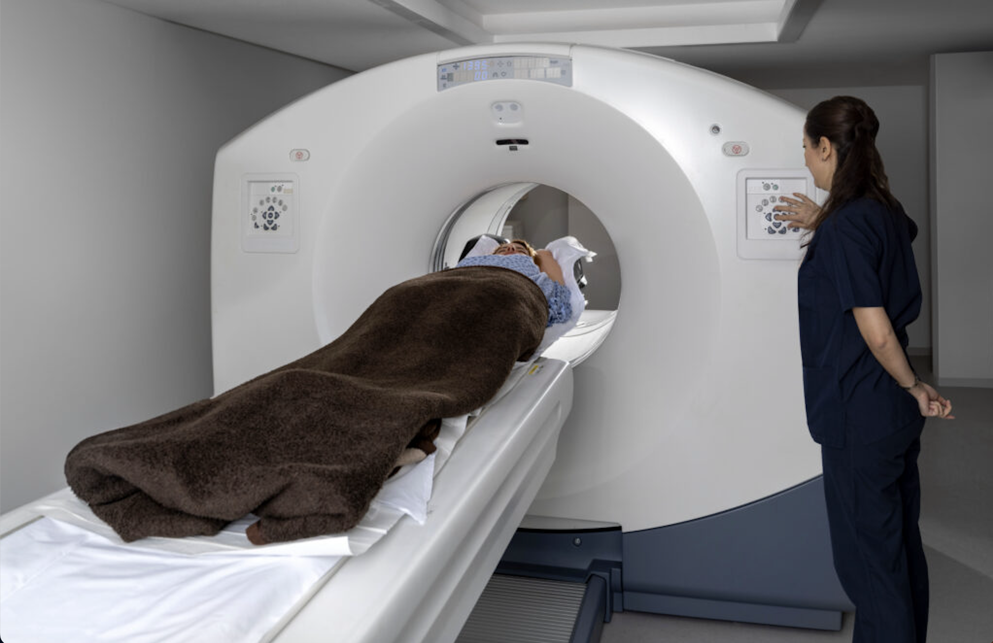
    When doctors want to see deeper into your body, PET-CT is a popular choice. It is a super powerful imaging tool that is widely used in diagnosis, staging, and monitoring of various diseases, particularly in oncology, cardiology, and neurology. It’s especially useful in detecting cancer, guiding treatment plans, and understanding conditions affecting the heart or brain. But how does it actually work — and why does it matter so much? Let’s figure it out.

    What is PET-CT exactly? The answer is quite straightforward. It is just PET plus CT! This means before we get into PET-CT, we have to learn about both PET and CT.

    Taking advantage of radioisotope signals, PET is an imaging method that enables the measurement of the change in metabolic activities and other physiological activities in vivo (Fowler & Volkow, 2022). All the radioisotopes are positron emitters that will emit positrons to keep them stable. Gamma rays will be produced and detected by sensors, which will be mapped. After millions of records, a clear map that provides information about the quantity and location of the positrons in the body will be produced (Basu et al., 2011).

    When interpreting the PET scanning image, the hot spots and the cold spots are what we need to focus on. Hot spots are the regions with bright colors (white, yellow, orange, and red). In these regions, the uptake rate of the radiotracer is higher, which indicates a higher rate of metabolic activity. Therefore, these regions will represent cancer, inflammation, or active regions of our body. Cold spots, on the contrary, represent dead tissue and reduced function that lower metabolism in that region (TerPogossian et al., 1980).

    Computed tomography (CT) is what we are more familiar with. It is a frequently used and widely available non-invasive imaging modality and has been applied in measuring the size of body tissues like bones, muscles, and brains. X-rays will be emitted from different directions, pass through the body, and be received by the detector, forming images of projections of the body in different directions. Through computer calculation based on the X-ray attenuation characteristics of the tissues within the voxel, sizes of targeted tissues will be measured (Mazonakis & Damilakis, 2016).

    With the basic knowledge of both PET and CT, it is easy to understand that PET-CT is just a combination of the two technologies. It can trace both the change in metabolism of the tissue and the size of it. On the other hand, the combination of these two technologies is quite revolutionary. One of the biggest limitations of a PET scan is its lack of spatial resolution and a clear anatomical reference frame. This defect hinders PET from accurately presenting the anatomic structure of lesion tissues, which negatively affects diagnosing diseases such as cancers and Alzheimer’s disease. Fortunately, CT addresses this problem. When combined, PET shows functional activity and CT shows structure, helping distinguish between normal and pathological signals. This fusion is especially important in cancer diagnosis, where identifying the exact location and stage of a tumor is critical for accurate treatment planning (Basu et al., 2011). With different kinds of radiotracers, PET-CT can be used in different aspects of imaging, including glucose metabolism, tumor proliferation, myocardial perfusion imaging, skeletal imaging, and brain imaging (Anand et al., 2009).

    With a lot of advantages, PET-CT is pervasively used around the world. Besides improving the accuracy of diagnosis, PET-CT also contributes to the early detection of certain cancers and the early detection of cancer recurrence, impacting patient management by assisting in defining potential candidates for curative surgery, planning the appropriate surgical or radiotherapy approach, and referring patients with unresectable disease to other therapeutic options (Israel & Kuten, 2007).

    It is clear that PET-CT is a life-saving tool for humans. However, there are still limitations we need to overcome. One of the major limitations of PET-CT as a cancer screening tool is its radiation exposure, which poses a potential health risk, especially when used routinely in healthy people. A standard PET-CT scan involves both a radioactive tracer and a CT scan. It is a combined radiation dose that can be significantly higher than most diagnostic imaging tests. This raises concerns about the long-term effects of repeated exposure, particularly in asymptomatic populations. Additionally, high cost and questionable cost-effectiveness further limit its practicality. PET-CT scans are expensive. When they detect abnormalities, they often lead to further testing and interventions, but many of them are unnecessary due to false positives or clinically insignificant findings. Moreover, PET-CT has limited sensitivity for certain cancers, especially those that are slow-growing or have low glucose metabolism, such as early-stage prostate cancer or some lung adenocarcinomas. These cancers may not show up clearly on PET scans, which could give patients a false sense of security and delay diagnosis. Overall, while PET-CT has diagnostic value in high-risk or symptomatic individuals, its limitations make it a controversial and potentially problematic tool for routine cancer screening in the general population (Schöder & Gönen, 2007).

    Despite its remarkable capabilities, PET-CT is not without challenges. While it has revolutionized the way we diagnose and monitor diseases, especially cancer, it is not yet ideal for general screening. The combined radiation dose, high cost, and limited sensitivity for certain low-activity cancers mean that routine use in healthy individuals may do more harm than good. False positives can lead to anxiety, unnecessary procedures, and added burden on healthcare systems. Still, in the hands of experienced clinicians and when used appropriately—particularly in high-risk or symptomatic patients—PET-CT remains an invaluable tool that can save lives by guiding targeted treatment decisions. As research continues and technology advances, there’s hope that PET-CT will become safer, more affordable, and even more precise. Until then, its greatest value lies in thoughtful, case-by-case application rather than widespread use for screening the general population. After all, smarter imaging leads to smarter care.


    References

    Featured image from Metastatic Trail Talk https://metastatictrialtalk.org/from-the-experts/pet-ct-scans/

    Anand, S., Singh, H., & Dash, A. (2009). Clinical Applications of PET and PET-CT. Medical Journal Armed Forces India, 65(4), 353–358. https://doi.org/10.1016/s0377-1237(09)80099-3

    Basu, S., Kwee, T. C., Surti, S., Akin, E. A., Yoo, D., & Alavi, A. (2011). Fundamentals of PET and PET/CT imaging. Annals of the New York Academy of Sciences, 1228(1), 1–18. https://doi.org/10.1111/j.1749-6632.2011.06077.x

    Fowler, J. S., & Volkow, N. D. (2022). Molecular Imaging: Positron Emission Tomography. Neuroscience in the 21st Century, 3297–3321. https://doi.org/10.1007/978-3-030-88832-9_149

    Israel, O., & Kuten, A. (2007). Early Detection of Cancer Recurrence: 18F-FDG PET/CT Can Make a Difference in Diagnosis and Patient Care. Journal of Nuclear Medicine, 48(1 suppl), 28S35S. https://jnm.snmjournals.org/content/48/1_suppl/28S.short

    Mazonakis, M., & Damilakis, J. (2016). Computed tomography: What and how does it measure? European Journal of Radiology, 85(8), 1499–1504. https://doi.org/10.1016/j.ejrad.2016.03.002

    Schöder, H., & Gönen, M. (2007). Screening for Cancer with PET and PET/CT: Potential and Limitations. Journal of Nuclear Medicine, 48(1 suppl), 4S18S. https://jnm.snmjournals.org/content/48/1_suppl/4S.short

    TerPogossian, M. M., Raichle, M. E., & Sobel, B. E. (1980). PositronEmission Tomography. Scientific American, 243(4), 170–181. JSTOR. https://doi.org/10.2307/24966439

  • Origami & Bio-inspired Design

    Origami & Bio-inspired Design

    You’ve probably seen pictures of origami before, or even folded some yourself! Origami is an ancient art form that originated in China in the 2nd century AD. It made its way to Japan around the 6th century, where it became an art for both recreation & decorating shrines. In the centuries since, it’s evolved into a complicated craft that produces beautiful art from a very simple medium—a single sheet of square paper. What you might not realize, however, is how closely origami is linked with science and math.

    The Mathematical Basis of Origami

    Whenever you fold a piece of paper, you’re performing an action that’s incredibly mathematical. The rules of geometry govern origami, and you can describe all possible folds in six axioms. These axioms tell you what kinds of geometric constructions & proofs are possible with origami and can be used to determine if a set of folds is even physically possible. If you want to learn more about these axioms, check out this webpage by origamist Robert J. Lang!

    Bio-inspired design is an interdisciplinary approach to design that takes inspiration from nature to design new technologies. You can combine the mathematical principles of origami with bio-inspired design to create novel robot inventions. Today, we’ll go through a few examples that span a wide spectrum of species!

    Worms, Snakes, and Other Slithery Ones

    Engineers are currently trying to develop better rescue robots that can navigate complex terrain. Worms, snakes, and other slithering & slinking creatures cross crazy (relative to them) terrain on a day-to-day basis, and are a great source of inspiration for scientists.

    Some robotics labs have taken inspiration from the movements of snakes, and have created robot origami snakes that slither around and replicate the moving patterns of their natural counterparts. These multi-segment robot prototypes have a versatile range of movement.

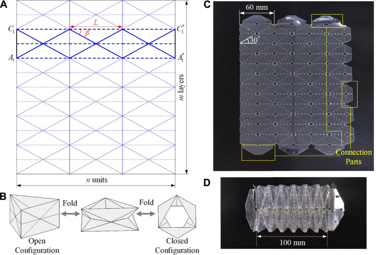
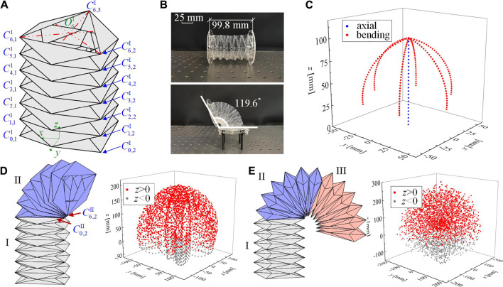
    Other labs have also mimicked the slinking movement of worms! Each of the segments in these worm-bots is made up of Yoshimura-Ori folds. You can see the crease pattern below in (A) of figure 1. The Yoshimura-Ori fold is a series of repeating triangles that can be collapsed into a cylindrical structure. They can be folded into an “open” or “closed” configuration which determines how compressed the unit is. A sequence of these folds is highly compressible & deformable while still maintaining its rigidity. It allows for a wide range of motion, which you can see in figure 2 of the movement of an earthworm inspired robot.

    Creaese diagram for yoshimura-ori folds, with diagrams of it expanded/contracted.
    Figure 1. Crease pattern of yoshimura-ori earthworm robot
    sequence of diagrams and photos showing the degree of motion of the worm robot
    Figure 2. Range of motion of yoshimura-ori folds

    The Yoshimura-ori structure allows the robot to mimic both the crawling motion that earthworms take as well as sharp directional turns. This technique holds a lot of promise for the future of all-terrain robots!

    Jellyfish & Aquatic Robots

    The land isn’t the only place origami is impacting robotics. The undulating motions that jellyfish swim with are both pretty and inspire researchers to develop new modes of underwater movement.

    Jellyfish are a type of organism called a medusa, which means that their body is umbrella shaped. Medusae are recognized to be the earliest multicellular organisms to have evolved muscular swimming, and while jellyfish generally float along ocean currents wherever they might go, they can use their hydrostatic skeleton to control vertical movement in a pinch. Prolate (long) medusae use jet propulsion to rapidly accelerate and avoid predators.

    They do this by contracting a water pouch in their bodies. The water expelled propels them in the opposite direction. They then expand their pouch and fill it with water again, ready to repeat for another burst of movement.

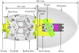
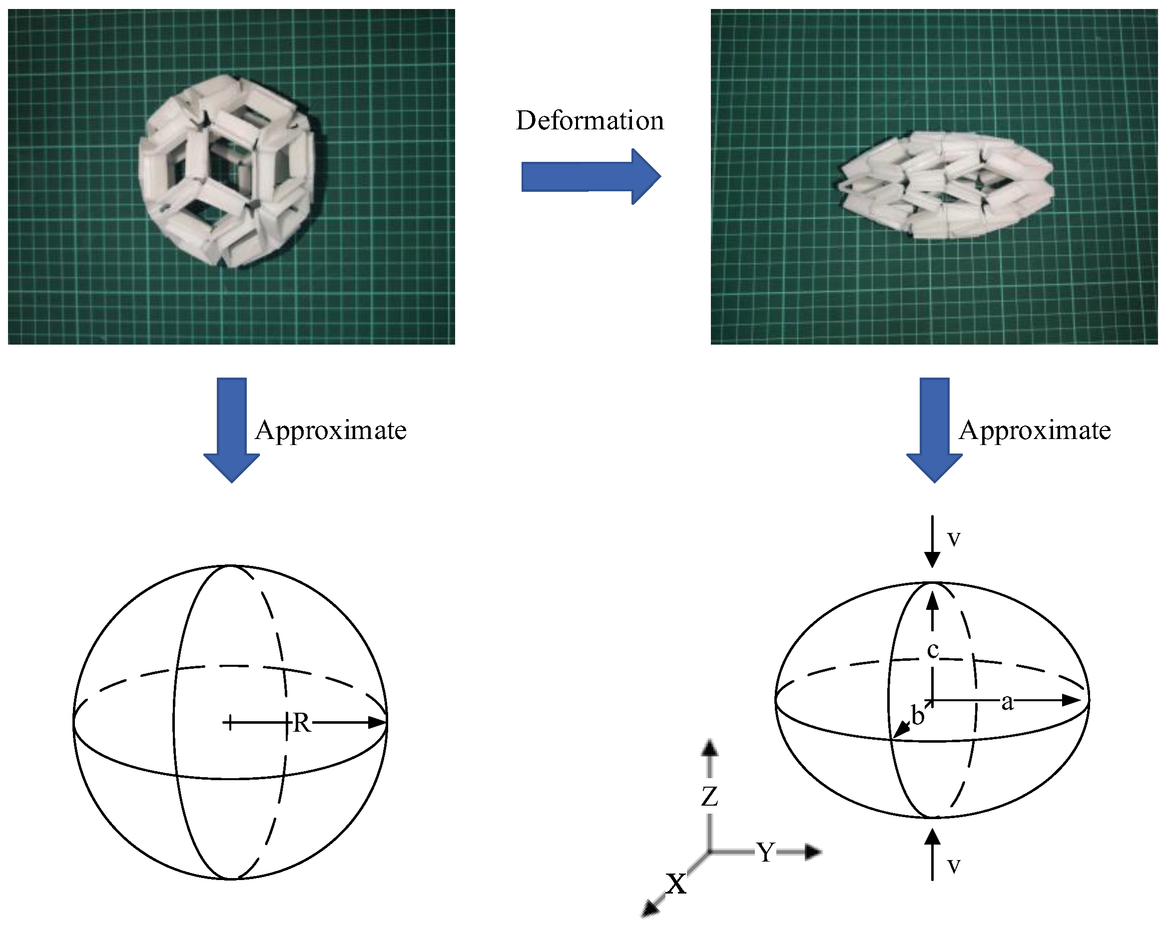
    Researchers are attempting to mimic this motion by combining soft-body robotics and origami engineering. The idea is that by creating a flexible origami polyhedral (a flexiball) they can motorize it in the same way medusae use jet propulsion underwater. The resulting flexiball they went with is known as a rhombic triacontahedron, and has 30 faces.

    Below is a picture of their 3D printed flexiball and how it changes shape after contraction.

    a before and after image of a 3d printed flexible triacontahedron being deformed
    Figure 3. Rhombic triacontahedtron flexiball

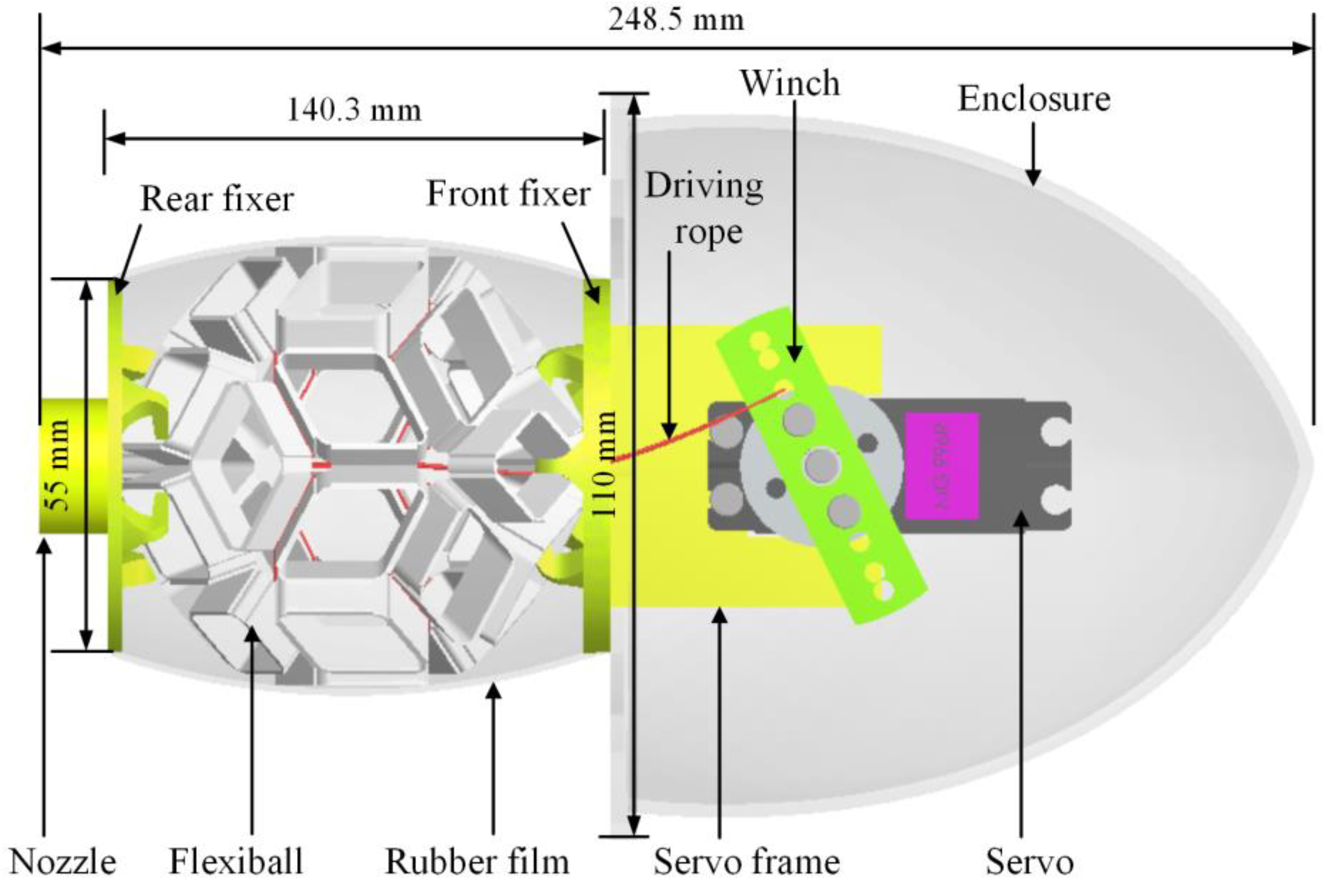
    The researchers then designed a lightweight, waterproof enclosure for the flexiball. The flexiball is contracted using three ropes connected to the grooves of the polyhedra’s edges.

    side profile diagram of jellyfish robot
    Figure 4. Side profile of jellyfish bot mechanisms

    Make your own!

    You don’t have to be an engineer to explore origami & science yourself! Try folding your own tessellation using the template below! Fold all the blue/dashed lines in one direction, and the red/dotted lines in the opposite direction.

    miura ori crease pattern
    Miura ori template! Try folding it yourself
    gif of miura ori fold expanding and contracting
    What your miura ori fold should do!


    Sources:

    Hu et al., “An Origami Flexiball-Inspired Soft Robotic Jellyfish.”

    Qiu et al., “Design and parameter optimization of a biomimetic jellyfish origami mechanism (BJOM) based on waterbomb tessellation.”

    Luo et al., “OriSnake: Design, Fabrication, and Experimental Analysis of a 3-D Origami Snake Robot.”

    Zhang et al., “Yoshimura-origami Based Earthworm-like Robot With 3-dimensional Locomotion Capability.”

    Schroeder, et al., “An electric-eel-inspired soft power source from stacked hydrogels.”

    Miura-ori crease pattern & GIF from Wikipedia Commons

  • Daguerreo-what? The Chemistry Behind Early Photography!

    Daguerreo-what? The Chemistry Behind Early Photography!

    Before smartphones and DSLRs were everywhere, photography was a physical, hands-on process. I’ll give you a quick overview of the chemistry behind the first type of photograph, the daguerreotype!

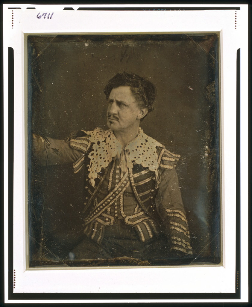
    Daguerreotype of Junius Booth, an actor, in theatrical garb
    Example of a daguerreotype

    Daguerreotypes were invented by Louis-Jacques-Mandé Daguerre in France in 1839. They consist of a picture developed on a silver-plated copper sheet. Each daguerreotype is a direct positive, meaning its image is created directly from the exposure without an intermediary negative. This contrasts with film, where your pictures are taken on a negative (a filmstrip) and then exposed as needed to create a positive. Because of this, the only way to replicate a daguerreotype is to rephotograph it, making each image incredibly unique!

    When preparing to take a daguerreotype, you would need to first polish a silver sheet until it was reflective like a mirror. Traditionally, to save resources photographers would use copper sheets plated with silver. Next, they would expose the plate to halide vapors to make the surface photosensitive Ag halides.

    When Louis Daguerre first invented the daguerreotype, he used iodine vapors. Iodine halides weren’t actually very photosensitive, and after a year of experimentation and innovation, people began to use more reactive halides such as chlorine and bromine as substitutes.

    The chemical formula for this step can be written down as:

    2Ag(s) + 1I2(g) à 2AgI

    2Ag(s) + 1Cl2(g) à 2AgCl

    2Ag(s) + 1Br2(g) à 2AgBr

    Once the plate is prepared, the photographer would set up the camera, pose their subject, and focus the lens. After their shot was composed, they would insert the plate into the camera’s plateholder, and begin taking the photograph. Because exposure times were so slow, instead of having a mechanical shutter, they would simply take the lens cap off to start the exposure and put it back on to stop it. While the plate is exposing, it would react with light:

    10AgX + 10hv à 2Ag5(s) + 5X2(g) (x being the halide)

    After the exposure is taken, portions of the plate that reacted to light consist of just Ag, while nonreactive ones remain AgX. At this point, the picture would still be invisible, and needs to be developed to be seen.

    Next, photographers would develop it using mercury vapor. This causes an amalgam (a mercury alloy) to form with the silver.

    11Ag5 + 45Hg(g) à 5Ag11Hg9 & 3Ag5 + 20Hg(g) à 5Ag3Hg4

    While this developed the image, it left the image in a highly reactive state. It could literally be wiped off by your hand! In order to fix the image to the plate, they would wash it with water and sodium thiosulfate. It would react with the thiosulfates to remove unreacted Ag halides and prevent the mercury amalgams from tarnishing.

    2Na2S2O3 + AgX à Na3[Ag8S2O3] + NaX

    This is where the traditional daguerreotype process finished! For display, they would be placed in a frame under glass to keep the picture safe. Further innovations to the technique included adding a layer of protective gilding & manually coloring the image.

    What makes the daguerreotype so cool is that it’s accurate down to the molecular level. Unlike digital cameras limited by pixels and film stock limited by crystal grain, daguerreotypes are direct exposures to light. If you look at a daguerreotype, you’ll notice they’re incredibly sharp images.

    Recently, there’s been a revival of traditional photographic techniques. Tons of practitioners have been experimenting with daguerreotype and other similar techniques such as wet-plate collodion, and many have been hosting workshops to introduce the public to them.

    Hopefully, this has sparked an interest in the daguerreotype. I’d encourage you to explore it more! It’s a really cool form of photography, and learning about it helps you appreciate the point-and-click cameras we take for granted today!

    Works Cited

    https://www.metmuseum.org/essays/daguerre-1787-1851-and-the-invention-of-photography

    https://medium.com/@mert.kty81/unveiling-the-past-daguerreotypes-and-the-legacy-of-mercury-fumes-a81f09398841

    https://daguerreiansociety.org/Making-a-Daguerreotype

    https://doi.org/10.3390/s23094341

    Picture credit Library of Congress

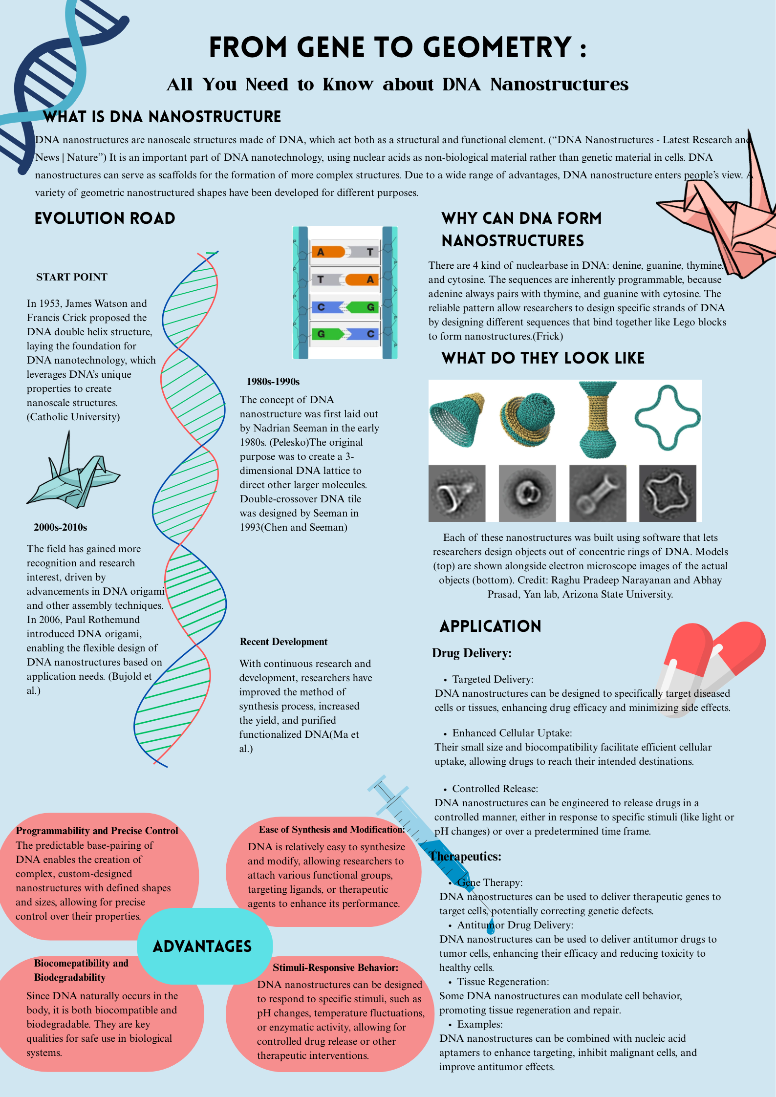
  • From Gene to Geometry : All You Need to Know about DNA Nanostructures

    From Gene to Geometry : All You Need to Know about DNA Nanostructures

    Works Cited
    Bujold, Katherine E., et al. “DNA Nanostructures at the Interface with Biology.” Chem, vol. 4, no. 3, Mar. 2018, pp. 495–521, https://doi.org/10.1016/j.chempr.2018.02.005.
    Catholic University. “School of Engineering.” The Catholic University of America, 2024, engineering.catholic.edu/research-and-faculty/faculty-profiles/eecs/hieu-bui/buiresearch/a-brief-introduction-to-dna-nanotechnology.html.
    Chen, Junghuei, and Nadrian C. Seeman. “Synthesis from DNA of a Molecule with the Connectivity of a Cube.” Nature, vol. 350, no. 6319, 1 Apr. 1991, pp. 631–633, www.nature.com/articles/350631a0, https://doi.org/10.1038/350631a0.
    “DNA Nanostructures – Latest Research and News | Nature.” Nature.com, Nature, 13 Jan. 2025, www.nature.com/subjects/dna-nanostructures. Accessed 3 Apr. 2025.
    Frick, Erin. “The Shape of Molecules to Come: A Q&a on Designing DNA Nanostructures for Biomedical Applications.” Phys.org, 6 Aug. 2024, phys.org/news/2024-08-molecules-qa-dna-nanostructures-biomedical.html. Accessed 3 Apr. 2025.
    Ma, Wenjuan, et al. “The Biological Applications of DNA Nanomaterials: Current Challenges and Future Directions.” Signal Transduction and Targeted Therapy, vol. 6, no. 1, 8 Oct. 2021, pp. 1–28, www.nature.com/articles/s41392-021-00727-9, https://doi.org/10.1038/s41392-021-00727-9. Accessed 18 Nov. 2021.
    Pelesko, John A. Self Assembly. CRC Press, 21 May 2007.
    Seeman, Nadrian C. “Structural DNA Nanotechnology: An Overview.” Methods in Molecular Biology (Clifton, N.J.), vol. 303, no. 15923682, 2005, pp. 143–166, www.ncbi.nlm.nih.gov/pmc/articles/PMC3478330/, https://doi.org/10.1385/1-59259-901-X:143. Accessed 25 Feb. 2022.