Category: Articles

  • What makes a figure interpretable?

    What makes a figure interpretable?

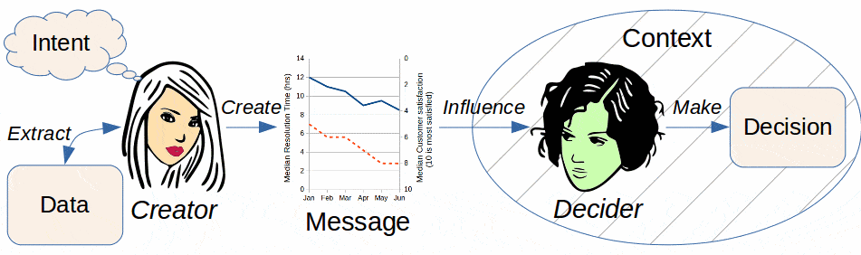
    Science communication is an often-overlooked but very important part of furthering the scientific process, and both researchers and publishers often use graphical means to display experimental results and data through figures, as presenting data this way is considered standard within reporting results and finding as it is easily interpretable for a vast array of audiences. However, as a neuroscience student, both involved with research in a computational cognition and neuroAI lab and involved with STEM communication as a communications assistant for an institutional research institute, an interesting nuance to researchers’ use of graphs and figures to portray their results, both statistically, as the results of an experimental analysis, and methodologically, which shows their experimental process, is that while we think that we know what cognitive mechanisms people are using while interpreting these figures, in reality, the neural mechanisms are, for the most part, completely unexplored.

    Inherently, almost anyone that you ask would believe that graph and figure comprehension is a visual task; however, research as recent as 2023 published by the National Eye Institute has shown that this might not be as true a heuristic – a mental shortcut or “cheat” that makes solving a problem easier – as initially thought. The National Eye Institute’s research showed that connections to the first region of the brain where visual information is processed, the primary visual cortex (V1), are rather small, accounting for between only 2 and 10 percent of neural connections that would be thought to inform us as to what we are perceiving. This is really interesting from a systems perspective in neuroscience, as this implies that what we are seeing only accounts for a relatively small portion of what we perceive. Therefore, in terms of graph and figure comprehension, the vast majority of neural connections are from other, non-visual,  regions of the brain, leading us to believe that our cognitive processing of graphs is largely a top-down process; a process where our understanding is heavily biased by prior experience and not what we are seeing in the moment.

    From our understanding of the cognitive processes of how we interpret graphs and figures, many different brain regions could theoretically be responsible for this task; until neuroimaging research is done, we will never know for certain. However, one region of the brain that is non-visual and could be responsible for and contribute to our ability to understand information embedded within graphs is the language network and its associated regions. The language network is a portion of the brain made up of many distinct regions, such as Broca’s and Wernicke’s areas, which are commonly known to be involved with speech generation and understanding, respectively, that neuroscientists know contribute to our linguistic abilities. This region of the brain, along with other associated regions such as the cerebral cortex, suggested by some recent research in the context of semantics (Ivanova et. al., 2025), might play a large role in our ability to understand and interpret graphs and figures.

    Additionally, research that has looked at large language models (LLMs), a form of artificial neural networks loosely modeled after how researchers understand the nervous system to process language, has interestingly shown that while really bad at processing graphs and figures, LLMs do technically use language to interpret and draw conclusions as to what they are looking at and pull from prior experience and reinforcement learning (Wang 2025).

    Overall, because of this, I believe that our ability to process and understand graphs and figures would rely on another cognitive mechanism, such as language or semantics, for example, where we can “think through” and connect what we are observing in a figure to information that we already know – so this inherent visual task, may not be as much about seeing, as it is about knowing. In general, rephrasing this question to What is the neural basis of data visualization comprehension? to explore The Semantics of Data Visualization is a really interesting idea that sits at the intersection of computational cognition, neuroAI, and STEM communication.  


    References

    Ivanova, A. A., Kauf, C., Gao, R., She, J. S., Kean, H. H., Goldhaber, T., Nieto-Castañón, A., Varley, R., Kanwisher, N., & Fedorenko, E. (2025). Semantic reasoning takes place largely outside the language network (Version 1) [Preprint]. bioRxiv. https://doi.org/10.64898/2025.12.07.692873

    National Eye Institute. (2023). Sparse, small, but diverse neural connections help make perception reliable, efficient. U.S. Department of Health and Human Services, National Institutes of Health. 

    Wang, H. W., Wang, Z., Xu, Y., Chen, Z., Wu, Y., Qu, H., & Hullman, J. (2025). How aligned are human chart takeaways and LLM-generated insights? IEEE Transactions on Visualization and Computer Graphics. https://doi.org/10.1109/TVCG.2024.10681139

     

  • What is Delusional Infestation?

    What is Delusional Infestation?

    What’s the weirdest psychiatric condition you’ve ever heard of? Odds are, this one is infinitely stranger. Delusional Infestation (DI) is a condition in which a person falsely believes that their skin is infested with parasites, insects, bacteria, fungi, or other harmful items (Campbell et al. 2019). They often experience symptoms such as itchy or tingling sensations, or even feel something crawling on or under their skin, which further convinces them that the infestation is real. They may bring “specimens” of the parasites to show their doctors and remain unconvinced when told that the specimens are harmless objects or that medical tests have found nothing physically wrong with their skin.

    The condition is also known as Delusional Parasitosis (DP), given that many people who have the condition attribute it to parasites. However, the terminology is changing in response to the fact that a wide variety of both living organisms and inanimate objects are blamed for symptoms (Bewley 2010).

    Once it has been established that the person is not actually infested, they may be diagnosed with Delusional Infestation if:

    • They have had the delusion for at least a month
    • Aside from the delusion, their lives are mostly unimpaired
    • Any mood episodes (mania or depression) that have occurred are brief compared to the duration of the delusion
    • Their symptoms are not better explained by another physical or mental illness, including schizophrenia, or by substance use* (Campbell et al. 2019)

    *If the case is primary DI; more on this in the next section

    Causes

    The cause of Delusional Infestation depends on the type of DI. Cases where DI is the only condition present are called primary DI. However, a person may also experience DI as a symptom of a different illness or due to the effects of certain drugs. In these situations, they are diagnosed with secondary DI because it is not the primary condition (Wikipedia Contributors 2019).

    The cause of primary DI is currently unknown, though it may have something to do with the neurotransmitter dopamine. The presence of unusually high amounts of dopamine in certain brain regions can create formication, the skin-crawling sensation experienced by many people with DI, and faulty dopamine activity has also been implicated in other psychotic disorders like schizophrenia. However, research on this topic is still developing, making it impossible to draw conclusions at this time (Wikipedia Contributors 2019).

    Secondary DI has quite a few potential causes. A vast range of physical medical conditions, from AIDS and hepatitis to traumatic brain injury and vitamin B12 deficiency, may cause delusions or crawling sensations (Reich et al. 2019, Wikipedia Contributors 2019). Consuming certain drugs, both legal and illegal, can also create similar effects. The side effects of some prescription drugs include DI, along with alcohol, cocaine, amphetamines, and cannabis (Campbell et al. 2019, Reich et al. 2019). Finally, other, more common mental conditions such as schizophrenia and depression may have symptoms that include DI.

    Treatment

    Thankfully, we know far more about treating DI than we do about its causes. Symptoms often respond well to antipsychotic medications, and in the case of secondary DI, they may vanish entirely when the person receives treatment for their primary condition (Campbell et al. 2019).

    Unfortunately, treatment of Delusional Infestation is often difficult, as by definition, people with DI believe that they have a physical illness, not a mental one. DI cases most often begin in the offices of dermatologists, parasitologists, and entomologists instead of psychiatrists. Upon being shown evidence that their condition is not physical (such as negative test results or proof that a specimen is ordinary detritus), a person may simply leave and go to a different doctor in an attempt to get the diagnosis they want. Recent discussions of DI among researchers and health care professionals have emphasized the importance of collaboration between a person’s doctors and maintaining a positive relationship with patients (Reich et al. 2019, Campbell et al. 2019). These measures can persuade some people with DI to agree to psychiatric treatment, but others may refuse to accept it no matter what.

    Potential Harms

    Many friends, relatives, and doctors who have seen the effects of Delusional Infestation become frustrated by their inability to persuade the affected person of the reality of their illness. At the same time, it is important to encourage DI patients to accept treatment, as the condition is not harmless. The most obvious problem caused by DI is the stress and mental suffering caused by the symptoms themselves – no one enjoys feeling like they have bugs inside their body! People with DI sometimes scratch or pick at their skin, potentially causing more issues. The symptoms and delusions may also cause problems at work and in social relationships. In rare cases, people with DI may harm themselves, other people, or pets in the course of attempts to get rid of the “infestation” (Shelomi 2026).

    A further danger exists in the form of people and organizations who, despite the seriousness of this condition, take advantage of vulnerable people with DI in order to gain money from them. There are multiple pseudoscientific diagnoses promoted in low-quality research and on the Internet that emphasize DI symptoms but cruelly encourage the delusion that the condition has a physical cause. Scary-sounding terms like “Morgellons Disease” and “Neurocutaneous Syndrome” are marketed to the public in order to promote the services of a sketchy doctor or sell a miracle cure (Shelomi 2026). Belief in these false diseases can prevent a person from getting real treatment or unnecessarily prolong their suffering by prompting them to chase after false cures for lengthy periods of time when they could have received help right away.

    Overall, Delusional Infestation is an interesting condition that exposes a number of phenomena related to the current state of science and medicine in our society. The need for more effective communication regarding rare conditions, the importance of trust between patients and doctors, and the prevalence of scammers that prey on vulnerable demographics are all important issues that deserve greater examination. DI demonstrates a complicated and strange relationship between our brains and bodies and exposes gaps in our knowledge of medicine and psychiatry. Hopefully in the future, it will get the attention it deserves in the form of more research into its causes and more effective treatments for those who suffer from its symptoms.


    Sources:

    Bewley, A.P., et al. “Delusional Parasitosis: Time to Call It Delusional Infestation.” British Journal of Dermatology, vol. 163, no. 1, 22 Feb. 2010, pp. 1–2, https://doi.org/10.1111/j.1365-2133.2010.09841.x

    Campbell, Elliott H., et al. “Diagnosis and Management of Delusional Parasitosis.” Journal of the American Academy of Dermatology, vol. 80, no. 5, May 2019, pp. 1428–1434, www.sciencedirect.com/science/article/pii/S0190962218330548, https://doi.org/10.1016/j.jaad.2018.12.012

    Reich, Adam, et al. “Delusions of Parasitosis: An Update.” Dermatology and Therapy, vol. 9, no. 4, 13 Sept. 2019, pp. 631–638, www.ncbi.nlm.nih.gov/pmc/articles/PMC6828902/, https://doi.org/10.1007/s13555-019-00324-3

    Shelomi, Matan. “Delusion or Dentistry: The Pseudoscientific Diagnosis of Neurocutaneous Syndrome | Skeptical Inquirer.” Skepticalinquirer.org, 4 Mar. 2026, skepticalinquirer.org/exclusive/delusion-or-dentistry-the-pseudoscientific-diagnosis-of-neurocutaneous-syndrome/

    Wikipedia Contributors. “Delusional Parasitosis.” Wikipedia, Wikimedia Foundation, 16 July 2019, en.wikipedia.org/wiki/Delusional_parasitosis

  • The Einstein’s Ghost Tour Video!

    The Einstein’s Ghost Tour Video!

    This semester, I had the opportunity to film and edit a video for our event w/ the Atlanta Science Festival! We hosted the Einstein’s Ghost Tour, where attendees walk through a tour and learn about the research and the untimely demise of four scientists throughout time: Archimedes, Carl Wilhelm Scheele, Joseph Crocé-Spinelli & Théodore Sivel, and Marie Curie. I don’t get the chance to do media for an event often, so I’m really glad I got the chance to. Videography is one of my favorite hobbies, and I wanted to challenge myself for this event! 

    With this video, I learned a lot of new techniques and skills. I’ve been learning to edit with Black Magic’s Davinci Resolve, which is one of the video-editing software that the film industry uses typically for color-grading. I wanted to practice and learn color grading, so I recorded in Log format, a camera setting that captures footage in a flat, desaturated way to preserve maximum dynamic color range and information. There are so many tutorials on color grading on YouTube! Because of that, there are also several ways to color grade, and it got really overwhelming for a newbie like me.

    One thing I found to be challenging was working with footage from a low-light setting. I can’t really color-grade too much or really fix the dark video in post-production. I think it’s hard for me to reach my fullest potential with the kind of footage I had to work with. Another challenge I faced was not getting any footage of the guests’ faces, which I think takes away from the recap video, because we couldn’t see their reactions or expressions to our actors’ lines. Hopefully, the final video is still good!

    I really enjoyed the trial-and-error of the entire experience: figuring out which colors to bring out, how to bring out certain colors without sacrificing others, which footage to keep and which to scrap, etc. I enjoy the creativity and open-ended aspect of video editing! It’s so much fun putting together various video footage to evoke emotions and tell a story. I hope I achieved that with my video!


  • Children Response to Conflicting Information

    Children Response to Conflicting Information

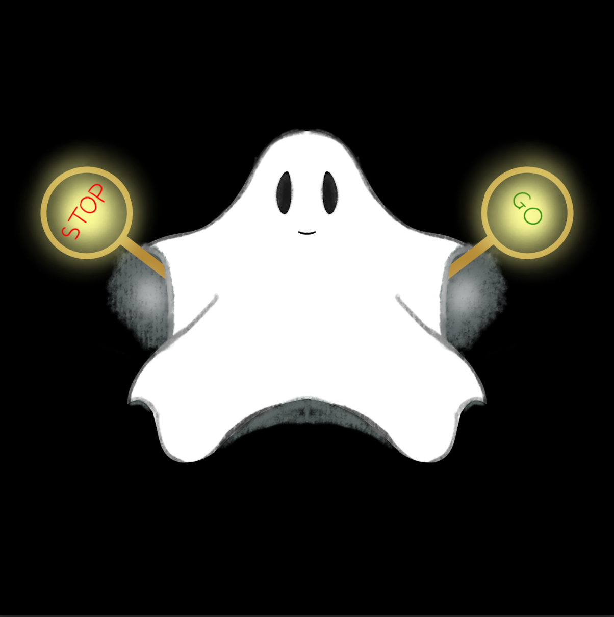
    <Featuring image is drawn by the author>

    Ghost Tour Observation

    Recently, the STEMComm VIP team took part in the Atlanta Science Festival in March 2026. We presented the Ghost Tour, featuring scientists who have died because of their inventions, passion, or major founding respective to their science field. I was part of the Zenith hot air balloon tragedy team as a NPC ghost actor. If you are interested, you can look up the three main characters: engineer Joseph Crocé-Spinelli, navy officer Théodore Sivel, and Gaston Tissandier. Two of them died on their flight into the death zone (high altitude), while Gaston survived to tell the story with the price of being deaf. 

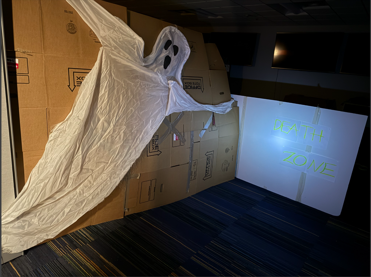
    Our team was assigned a big room. As guests come in from the door, they will be blocked by a big board sign of “death zone”, suggesting the death zone is ahead. The guide of the tour – Einstein, will lead them to the sign and leave them there. In the meantime, the NPC ghost will sneak in between the tour group, surprise the guests, and walk them past the sign, closer to the actual scene featuring the actors.

    <Picture is taken by the author>

    However, a small issue arised.There was a mix of reactions when the NPC ghost took the lead to walk over the Death Zone sign. Most people stayed still behind the sign, some hesitated to walk over, and very few actually followed the ghost.


    Mixed Behaviors to Conflicting Information

    Apparently, at first, the guide – Einstein, told the guests to not cross over the death zone. Then, the ghost NPC told them “follow me” as the ghost moved past the sign. Even with the actual dead scientists signaling and inviting the guests to come closer, it takes some time for them to actually move.

    • First information: Do not cross the death zone – Guide Einstein said
    • Second information: Follow me (crossing death zone) – NPC ghost moved
    • Third information: Come closer – Dead scientists in the death zone, with obvious hand signals to come closer

    There were different tour groups. The reactions are also mixed between these groups. Groups with more children tend to follow the NPC ghost at first, with young children being the first to move. In groups with more adults, the hesitation is very obvious. Even when children start walking over, they are stopped by the guardians. It takes the three actors’ effort to invite them over.


    How Children Reaction to Conflicting Sources

    Lili Ma and Patricia Ganea (2009) suggest that children often learn about the world through direct observation, but their knowledge can also be acquired through others’ testimony. This is validated as children often trust what they see, but they also recognize adults as trustworthy sources. Furthermore, some experiments suggest that children might accept false testimony that contradict their first-hand observation. This aligns with the classic Asch study where adults’ judgement was influenced by the social pressure and testimony of others, even though their visual perception suggests otherwise (Ma & Ganea, 2009). This explains why young children who were ready to walk over stopped along with their guardians. Parents are the more trustworthy source of information.

    In addition, the three studies done by Ma and Ganea (2009) confirms that children can outweigh one source of information more than another. This aligns with another study by Baer and others (2025), suggesting that children endorse confident informants over hesitant ones when faced with conflicting testimony. The intention of one ghost to walk over might not have been as clear compared to a group of familiar adults staying in place. On a side note, in the age of the Internet, even if the confident informants previously gave false testimony, it is unlikely they will lose credibility. This is true for both children and adults as they still follow Einstein the guide after the hot air balloon room (Guerrero et al., 2020). 

     Interestingly, studies also show that young children can accept both sources, even if there is a contradiction (Ma & Ganea, 2009). Over time, children learn to distinguish reliable sources over misleading testimony. This is crucial to their development of selective trust and skepticism. At one point, children can conclude new understanding of the situations based on contradiction sources. This might happen around the age of 8 (Baer et al., 2025). With the second piece of information contradicting the first (and their trustworthy informants), then the last information supporting the second, the young guests were able to resolve the tricky situation and take the lead to start the scene.


    Afterthought

    Adults tend to apply the situation in their thought process. Given it was a Ghost Tour, they might assume to not just listen to the wandering ghosts. In addition, did Einstein’s credibility as the guide play in his favor? Einstein was the original source of information for the guests.  Guests might assume the ghosts are just acting out the script. What would you do in their shoes?


    References:

    Baer, C., Engelmann, J. M., & Kidd, C. (2025). Children use the relative confidence of people with conflicting perspectives to form their own beliefs. Developmental Science, 28(4). https://doi.org/10.1111/desc.70027

    Guerrero, S., Sebastián-Enesco, C., Morales, I., Varea, E., & Enesco, I. (2020). (in)sensitivity to accuracy? children’s and adults’ decisions about who to trust: The teacher or the internet. Frontiers in Psychology, 11. https://doi.org/10.3389/fpsyg.2020.551131

    Ma, L., & Ganea, P. A. (2009). Dealing with conflicting information: Young Children’s reliance on what they see versus what they are told. Developmental Science, 13(1), 151–160. https://doi.org/10.1111/j.1467-7687.2009.00878.x 


  • The Clock Within: Neurospora crassa

    The Clock Within: Neurospora crassa

    The Clock Within: Neurospora crassa

    What is the science behind the clock?

    How did the first person just acknowledge that “Oh it’s 12:00 p.m.”? 

    Wouldn’t it surprise you to realize that your body already knows time?

    The human body has a natural mechanism to tell the precise time without any measuring device.

    Even if a person was kept in complete darkness for many days, they won’t forget the time. They will still experience sleepiness and wakefulness but the cycle will gradually change without any synchronization with sunlight.

    This question also aroused curiosity in the minds of researchers, but none could have predicted that bread mold would provide the solution.

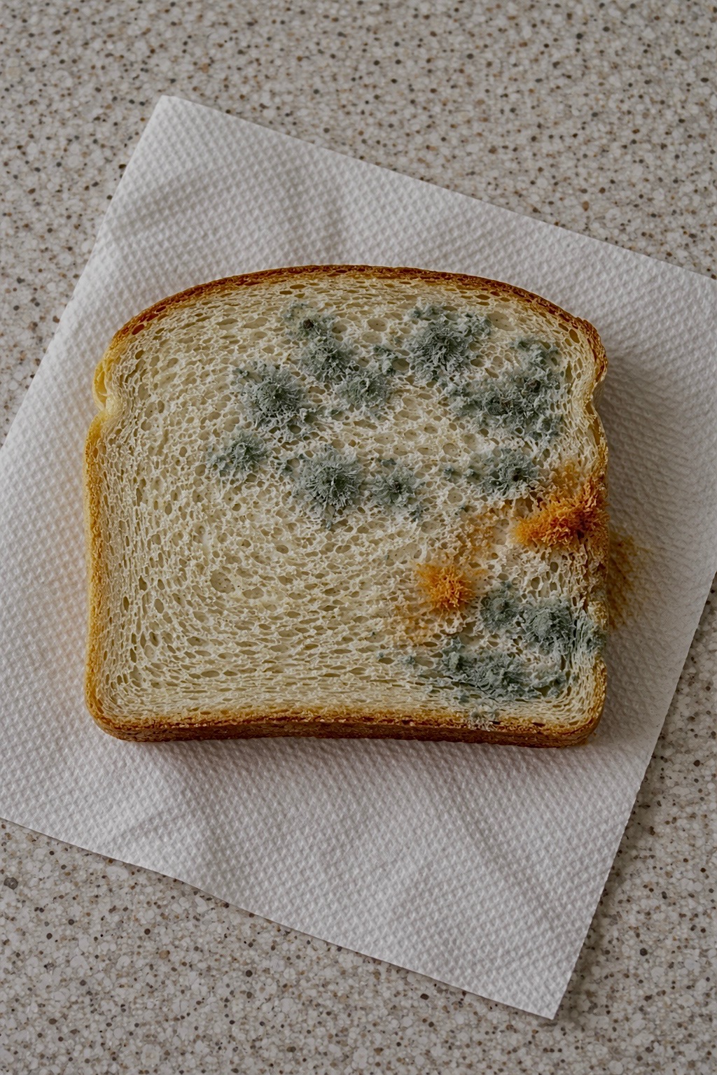
    In 1959, scientists began studying an orange bread mold by the name of Neurospora crassa.

    I even did a test like this myself, leaving bread out for several days just to observe what happened to the mold (seen below).

    Somehow, this small organism allowed scientists to understand how living things, including humans, keep track of time using a biological clock.

    It did not seem too important at first. But as soon as they took it into their laboratory, it did something rather unusual. They placed the fungus in complete darkness, with no sunlight, not even a single outside signal. The fungus could not tell what time it was. Even then, it still grew in certain patterns (Castro-Longoria et al., 2010). It repeated in a consistent cycle, as though the organism already knew what a day looked like.

    This insight changed the perspective on time.

    Scientists such as Jay C. Dunlap and Jennifer J. Loros concluded that organisms do not just respond to time but keep an internal clock that runs in cycles (Castro-Longoria et al., 2010). Such a system came to be called a circadian rhythm, meaning “biological rhythm occurring every 24 hours.”

    Scientists later discovered that this clock runs through interactions among genes and proteins in a cycle. A vital part of this cycle includes a gene whose job is to create a certain type of protein named FRQ (frequency) (Baker et al., 2011).

    When the FRQ protein accumulates in the body, it stops being produced. When this protein degrades, production resumes. Through this process, a continuous cycle is formed, allowing time to be regulated. This is known as a negative feedback loop, where a system turns itself on and off (Baker et al., 2011).

    This system is not simple. However, there are other genes, which help the organism to sense light. These are collectively referred to as the “white collar” genes (WC-1 and WC-2). They work together in a cycle and are almost always around 24 hours in duration despite any change in the environment.

    Humans function with a similar system.

    The human brain has an intricate structure called the Suprachiasmatic Nucleus (SCN), which serves as the timekeeper for the organism, regulating physiological processes like sleep, hormone secretion, and body temperature. Just like in fungi, the circadian rhythm is regulated via protein-gene feedback loop mechanism.

    Circadian rhythms have been observed across different organisms, suggesting that the biological mechanism for keeping time is ubiquitous and therefore fundamental for any living thing (Aramayo & Selker, 2013).

    This was actually quite simple. Our internal clock had always been around.It was only when Neurospora crassa revealed itself that we realized its existence.

    References:

    Baker, C.L., Loros, J.J., & Dunlap, J.C. (2011). The circadian clock of Neurospora crassa.
    https://pmc.ncbi.nlm.nih.gov/articles/PMC3203324/

    Aramayo, R. & Selker, E.U. (2013). Neurospora crassa, a Model System for Epigenetics Research.
    https://pmc.ncbi.nlm.nih.gov/articles/PMC3783048/

    Case, M.E., Griffith, J., Dong, W., Tigner, I.L., Gaines, K., Jiang, J.C., Jazwinski, S.M., Arnold, J.,  (2005). The aging biological clock in Neurospora crassa.
    https://pmc.ncbi.nlm.nih.gov/articles/PMC4228622/

    Castro-Longoria, E., Ferry, M., Bartnicki-Garcia, S., Hasty, J., Brody, S. (2010). Circadian rhythms in Neurospora crassa: Dynamics of the clock component frequency visualized using a fluorescent reporter
    https://www.sciencedirect.com/science/article/pii/S1087184509002242

  • Circus Sea Slugs: How clown nudibranchs are like real-life Pokémon

    Circus Sea Slugs: How clown nudibranchs are like real-life Pokémon

    The Pokémon “Shellos” and its evolution “Gastrodon” (whose pink West Sea variant is one of my favorite Pokémon) resemble a sea slug. Don’t just take my word for it, though—in their Pokédex entries, Shellos and Gastrodon are referred to as the “Sea Slug Pokémon”. The Pokémon Goomy is referred to as a Mollusk Pokémon in many languages, and although sea slugs are a type of mollusk, the resemblance is less apparent. The team behind the games really didn’t have to change much from their real-life inspiration, though, as sea slugs are some of the most unreal-looking creatures in our oceans. As Helen Sullivan writes about sea slugs, which are also known as nudibranchs, in the Smithsonian Magazine, “It is really, really difficult not to compare them to Pokémon characters” (2024).

    Nudibranchs belong to the class Gastropoda, which falls under the phylum Mollusca (mollusks). The term sea slug is used to broadly refer to 11 different subgroups under Gastropoda that do not have hard outer shells. True nudibranchs account for only one of these subgroups, but the others are sometimes also referred to as nudibranchs for convenience (Davey & Willan, 2020). 

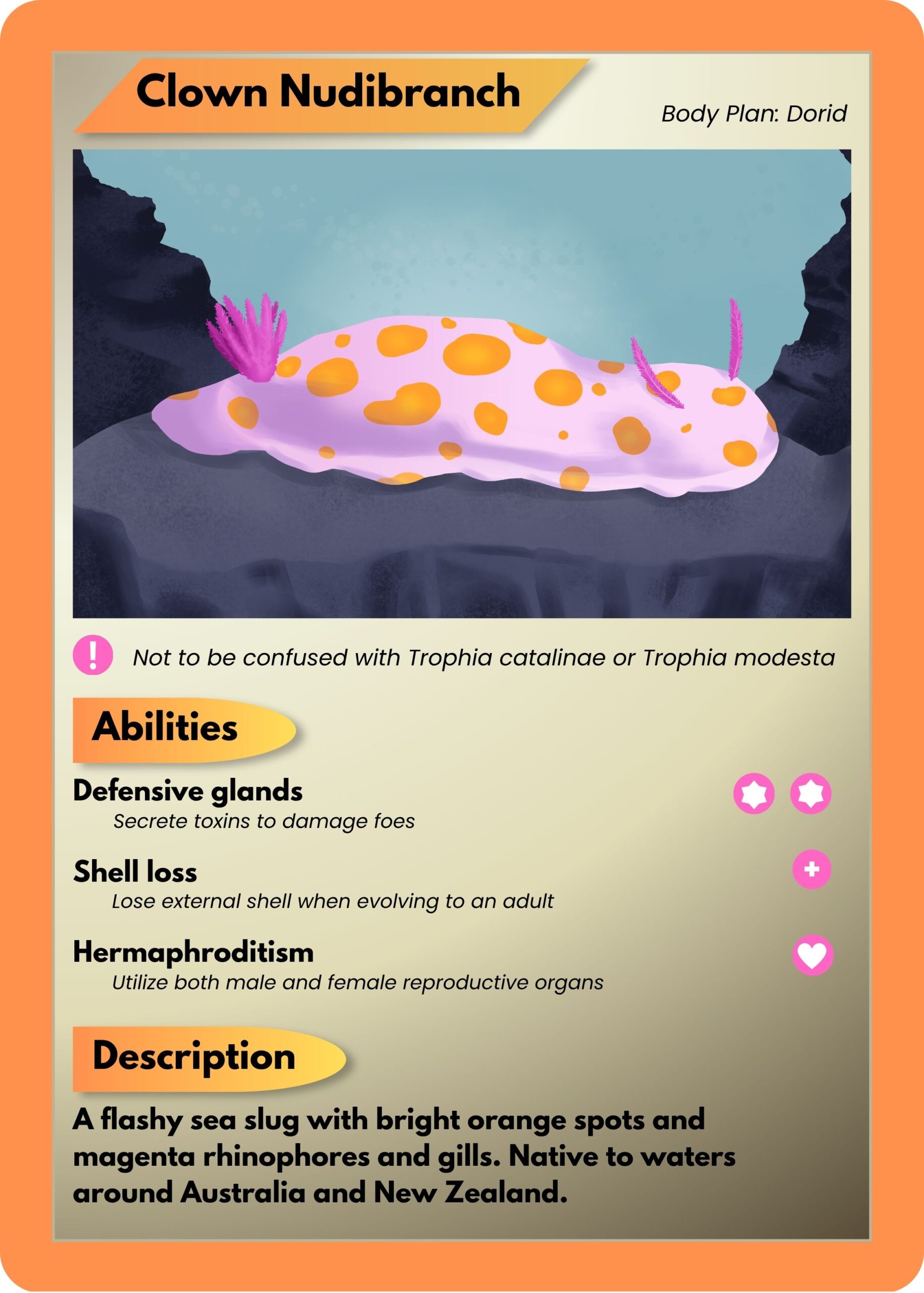
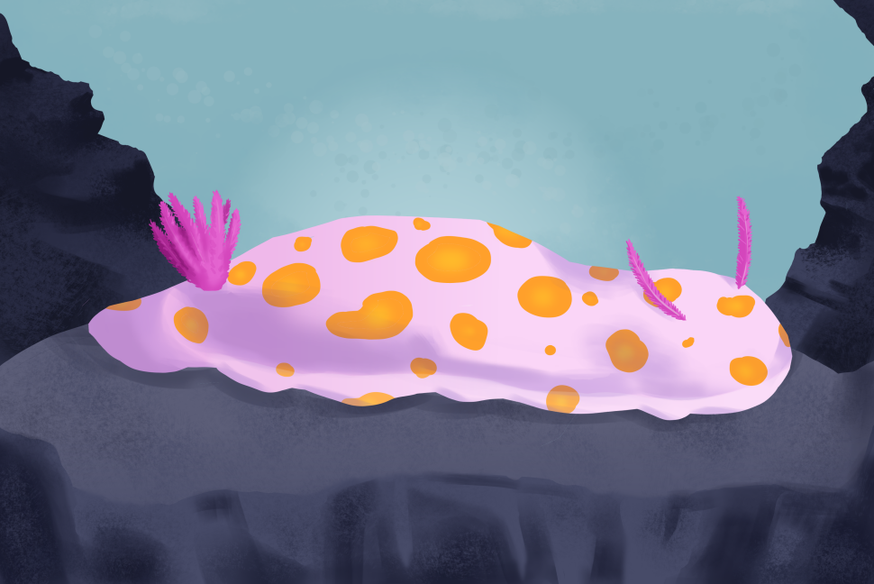
    When reading about species of nudibranch, one caught my eye for its name and bright orange coloration: the clown nudibranch. Even the name makes them sound like some sort of Pokémon! They are quite small—around 3 cm on average for adults—but their orange spots would probably make them hard to miss. The name, appearance, and distinctive characteristics of this creature gave me the idea to make a faux Pokémon card for the species, whose scientific name is Ceratosoma amoenum.

    Illustration/Graphic Design by Priyanka Singh

    Dorid

    Nudibranchs can have a few different body plans that differ in shape, gills, and more. C. amoenum has a dorid nudibranch body plan, which means it has two protrusions on its front end and a bundle of protrusions on its back end. The two protrusions that resemble antennae are called rhinophores while the bunch of protrusions on its backside are actually its gills! This distinctive characteristic is where nudibranchs get their name from. Nudibranch means “naked gills” (Sullivan, 2024).

    Not to be confused with…

    When researching for this article, I found that there are other nudibranch species with a similar appearance and common name as C. amoenum. Trophia catalinae also has a white body with orange spots, although its spots are raised (known as “tubercles”), and its rhinophores and gills are orange rather than magenta. It’s referred to by a similar name: “sea clown trophia” or simply “sea clown”, so you can see where the confusion might arise. Trophia modesta is a species that was recently distinguished from Trophia catalinae in a 2020 study, and it is commonly known as the “modest clown dorid” (Jung et al.). Unsurprisingly, it looks more similar to T. catalinae than it does to C. amoenum, although its body is more translucent than T. catalinae’s. Still, an amateur nudibranch enjoyer may get these species confused at first glance.

    Defensive glands

    Nudibranchs often contain chemical defenses within their bodies. A protective tissue called the mantle contains glands that secrete the toxins (Davey & Willan, 2020). Like other nudibranchs, our clown friend doesn’t produce these toxins on their own—they obtain them from their diet, which consists primarily of sponges (Winters et al., 2018). This chemical defense is critical for the soft, shell-less nudibranchs.

    Shell loss

    Speaking of not having a shell, both the English and Japanese name for Shellos mean “lost shell”: Karanakushi in Japanese, with kara meaning “shell” and nakushi being derived from nakushite (without) or nakusu (to lose); and “shell loss” (Shellos) in English (“Shellos”, 2026). The name origin references one of the nudibranch’s most defining characteristics. Gastropods have an external shell in their larval stage, although members of order Nudibranchia lose it before they reach adulthood, with C. amoenum being no exception (Davey & Willan, 2020). 

    Hermaphroditism

    As members of the Gastropoda class, sea slugs are hermaphrodites. Each individual has both male and female reproductive organs. I know what you may be wondering, but no, they can’t impregnate themselves, although they often donate and receive sperm simultaneously with another individual. Some nudibranchs even shed their penises after intercourse and gain a new one soon after (Sekizama et al., 2013). Talk about freaks of nature….

    I considered designing a creature based on the clown nudibranch for my fake Pokémon card, but why would I when the real thing looks like that? I’ll leave the creature designs to the Pokémon team for now. I hope you’ve come to see that sometimes reality can be stranger than the fiction it inspires, and that our world is full of amazing yet often overlooked creatures like the nudibranch.

    Illustration by Priyanka Singh

    References

    Davey, N., Willan, R.C. (2020). Super sea slugs: a guide to the sea slugs of New Zealand. (Version 1). National Institute of Water and Atmospheric Research (NIWA). 

    Gastrodon. (2026). Bulbapedia. Retrieved April 17, 2026, from https://pokemondb.net/pokedex/gastrodon 

    Jung, D., Gosliner, T.M., Choi, T., Kil, H., Chichvarkhin, A., Goddard, J.H.R., Valdés, A. (2020). The return of the clown: pseudocryptic speciation in the North Pacific clown nudibranch, Triopha catalinae (Cooper, 1863) sensu lato identified by integrative taxonomic approaches. Marine Biodiversity, 50, 84. doi.org/10.1007/s12526-020-01107-2 

    Sekizama, A., Seki, S., Tokuzato, M., Shiga, S., Nakashima, Y. (2013). Disposable penis and its replenishment in a simultaneous hermaphrodite. Biology Letters, 9(2). doi.org/10.1098/rsbl.2012. 1150 

    Shellos. (2026). Bulbapedia. Retrieved April 17, 2026, from https://pokemondb.net/pokedex/shellos 

    Sullivan, H. (2024, May). Dive Into the Exotic World of Nudibranchs, the Spectacular Slugs of the Sea. Smithsonian Magazine. www.smithsonianmag.com/science-nature/dive-exotic- world-nudibranchs-spectacular-slugs-sea-180984013/

    Winters, A.E., White, A.M., Dewi, A.S., Mudianta, I.W., Wilson, N.G., Forster, L.C., Garson, M.J., Cheney, K.L. (2018). Distribution of Defensive Metabolites in Nudibranch Molluscs. Journal of Chemical Ecology, 44, 384-396. doi.org/10.1007/s10886-018-0941-5 

  • Tech is in Bloom for Spring!

    Tech is in Bloom for Spring!

    It’s currently spring at Georgia Tech, which means tons of flowers are in bloom. Around the end of March and beginning of April, students, staff, and visitors can all take a walk around campus to take in the natural beauty of Georgia Tech. 

    Here are some of the plants that they may run into:

    Piedmont Azaleas: 

    One of the first few flowers that one might notice around Tech are the Piedmont Azaleas, which often grow in bushes on the walkway up to the Howey Physics building.

    The Piedmont Azalea, also known as Rhododendron canescens, is a flower native to Georgia. These azaleas bloom early in the spring, from mid-February through March (Wade, Braman, & Woodward, 2022).

    They tend to grow in the earlier parts of spring and produce a sweet, musky scent in the air. (Yadav et al., 2019). These flowers often attract a number of pollinators, including butterflies, bees, and hummingbirds. 


    Flowering Dogwood:

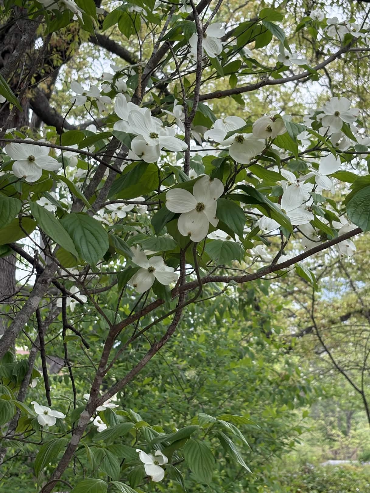
    Outside the Kendeda building also lies flowering Dogwood trees, which are known as the Cornus florida.

    These trees are often planted throughout Georgia as ornamental trees. This means that the trees are mainly planted for display rather than a more practical function. 

    However, the species can be extremely beneficial to the environment. Dogwood trees produce leaf litter that decomposes rather quickly, which can improve the soil quality (USDA). Additionally, the plant is an extremely important part of calcium cycling. Plants need calcium to survive, as it is extremely important to the structure of cell walls and plasma membranes. Flowering dogwood trees draw calcium from the soil, cycling it into a more usable form for other plants. (Borer, Sapp, & Hutchinson, 2013).


    Philadelphia Fleabane:

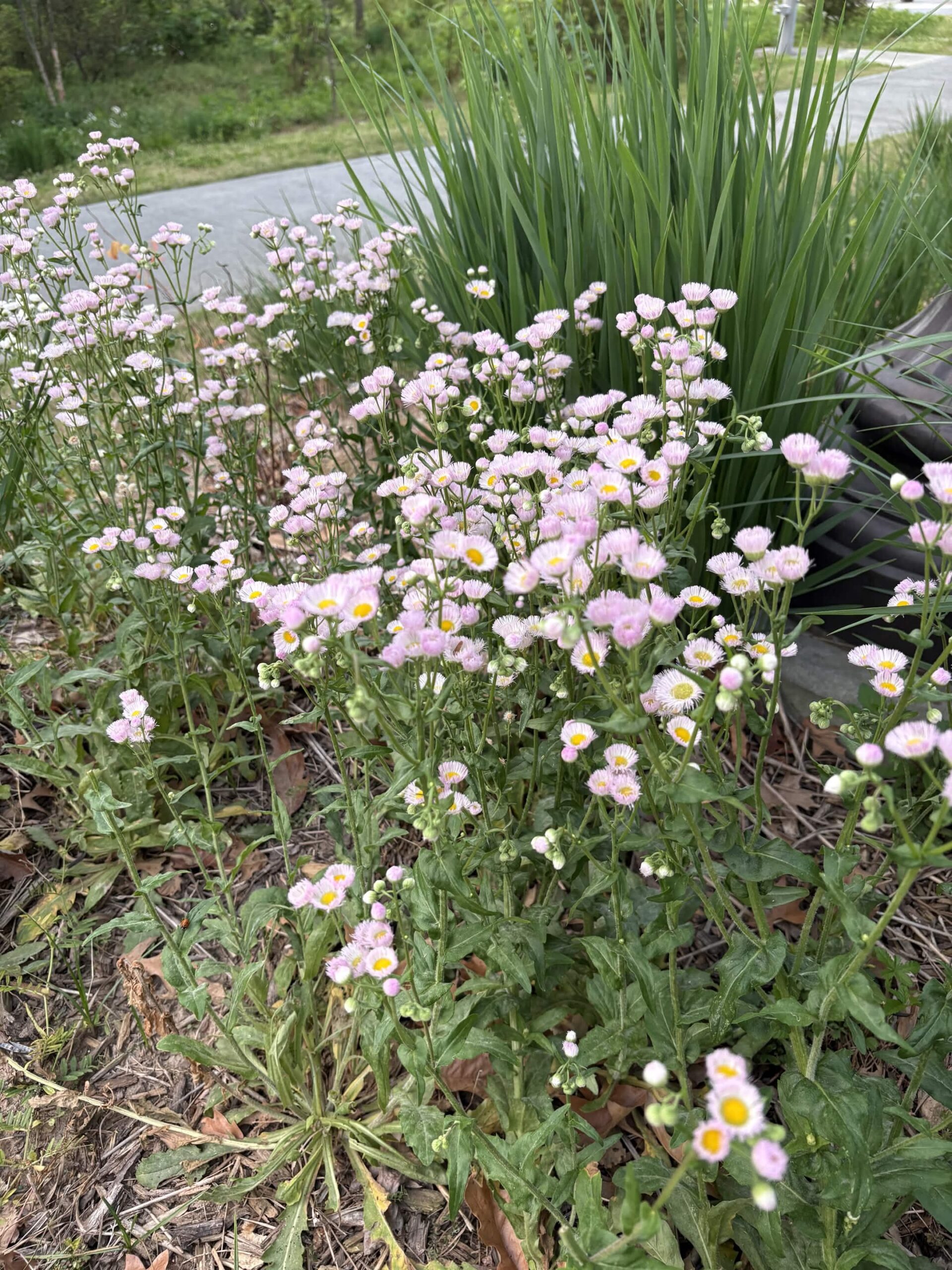
    Around the Center Street South apartments are the Philadelphia Fleabane, which are also known as the Erigeron philadelphicus.

    These are common wildflowers that grow in moist regions of the United States. They are often considered as weeds if they are not managed properly. This does not mean that they are invasive, but rather that the plant can grow in unwanted spaces if they are not managed properly. 

    They most commonly bloom during the spring. The flowers will remain open throughout the day time and close back up during the night.

    Throughout history, these flowers have had a number of uses, especially in Native American populations. The Philadelphia Fleabane has been made into tea, burned as incense, or simply chewed to treat the common cold in the past (USDA). 


    Oakleaf Hydrangea:

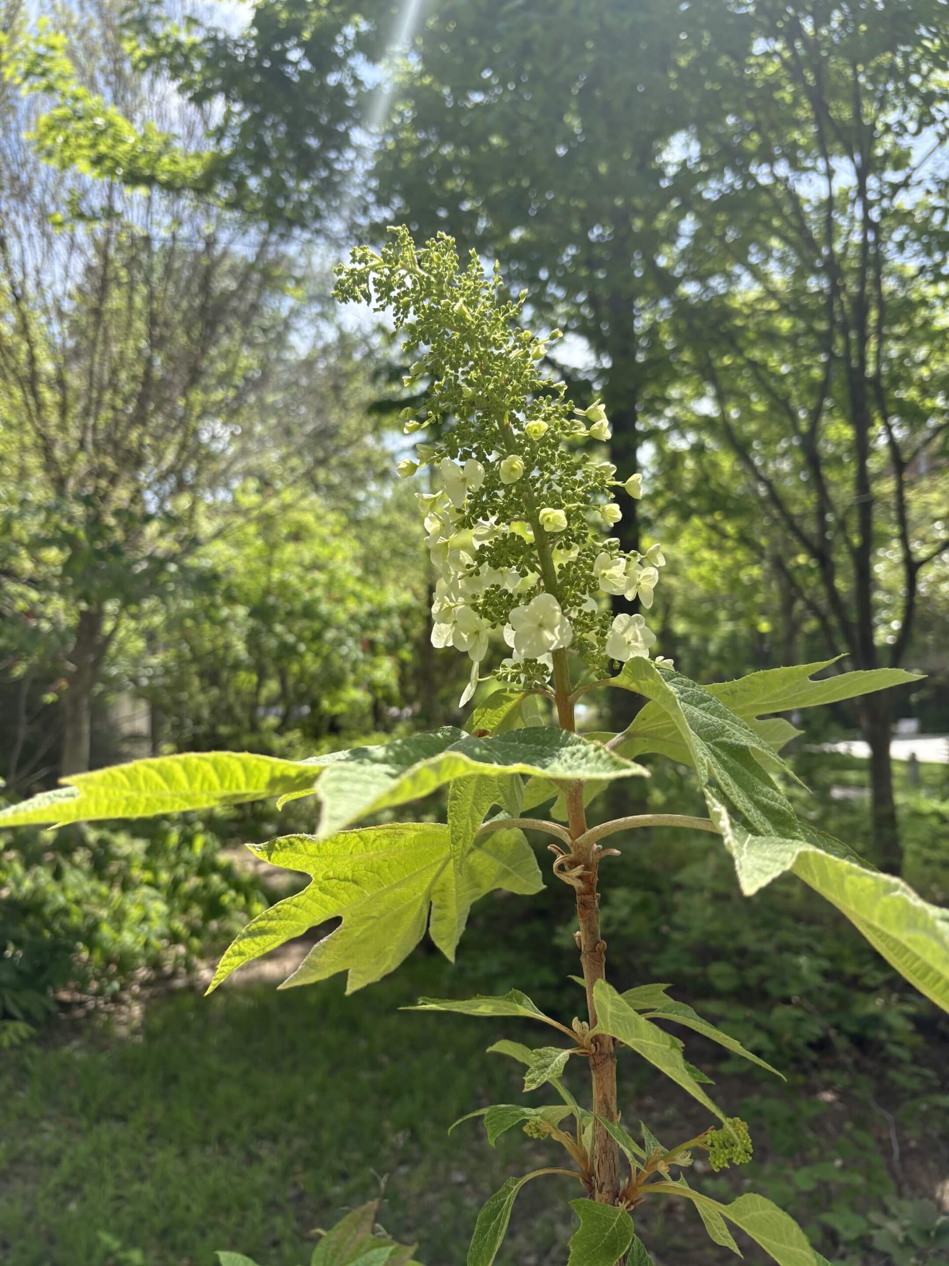
    Finally, we have the Oakleaf hydrangeas, which are generally grown in shrubs near the Klaus and Van Leer buildings towards late spring or early summer. These are also known as Hydrangea quercifolia, and they can be found blooming throughout campus. The flowers are native to the southeastern parts of the United States, where they begin to bloom in the early parts of summer.

    The plants grow in shrubs, with many cone-like structures that eventually bloom with cream-colored flowers. They are more known to be planted for ornamental purposes.

    Although the hydrangea pictured is native to Georgia, there are other species of Oakleaf hydrangeas that were specifically bred for their looks. For example, universities from multiple states in the southeast came together to cultivate the Ruby Slipper Oakleaf. It is a hybrid that starts off white just like the flower pictured and later turns a deep red color as it ages (Reed, 2010).


    Works Cited:

    Borer, C.H., Sapp, S.G. & Hutchinson, L.H. (2013). Flowering dogwood (Cornus florida L.) as mediator of calcium cycling: new insights are revealed by analysis of foliar partitioning. Trees 27, 841–849 https://doi.org/10.1007/s00468-012-0838-9 

    Georgia Native Plant Society. (n.d.). Piedmont Azalea (rhododendron canescens). GNPS https://gnps.org/plant/piedmont-azalea-rhododendron-canescens-2/ 

    Reed, S. M. (2010). ‘Ruby Slippers’ and ‘Munchkin’ Oakleaf Hydrangeas. HortScience, 45(12), 1908–1909. https://doi.org/10.21273/HORTSCI.45.12.1908 

    United States Department of Agriculture National Resources Conservation Service, USDA. (n.d.). PHILADDELPHIA FLEABANE Erigeron philadelphicus L.. https://plants.sc.egov.usda.gov/DocumentLibrary/plantguide/pdf/cs_erph.pdf 

    United States Department of Agriculture National Resources Conservation Service, USDA. (n.d.). FLOWERING DOGWOOD Cornus Florida L. https://plants.sc.egov.usda.gov/DocumentLibrary/plantguide/pdf/pg_cofl2.pdf 

    Wade, G. L., Braman, K., Woodward, J. W., (2022) Selecting and Growing Azaleas. UGA Cooperative Extension Bulletin 670. https://fieldreport.caes.uga.edu/wp-content/uploads/2025/08/B-670_6.pdf 

    Yadav, L. K., McAssey, E. V., & Wilde, H. D. (2019). Genetic Diversity and Population Structure of Rhododendron canescens, a Native Azalea for Urban Landscaping. HortScience, 54(4), 647–651. https://doi.org/10.21273/HORTSCI13840-18