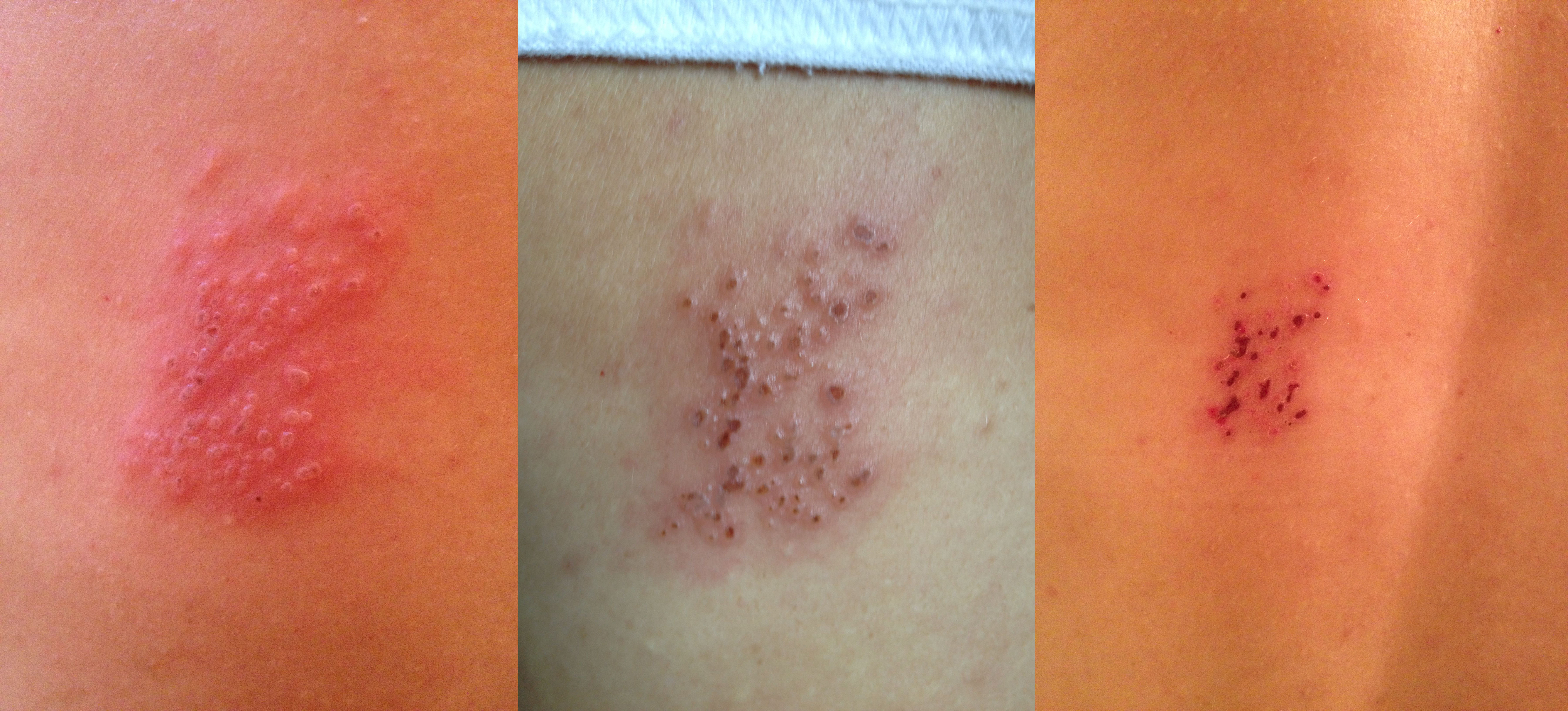
Ultraviolet radiation: what you can’t see can still hurt you

A few weeks ago, a friend of mine showed me an article on MSN about a man whose face was obviously more wrinkled on one side than the other. Mr. McElligott, the gentleman featured in the article, drove a delivery truck for 28 years, exposing the left side of his face to the sun for several hours each day. I have heard several times throughout the years about the importance of sun protection, but hadn’t ever dwelled on the topic for much time. Once I read about Mr. McElligott, though, I was quite intrigued and started thinking about the whys and hows of sun damage, UV radiation, and wrinkles.
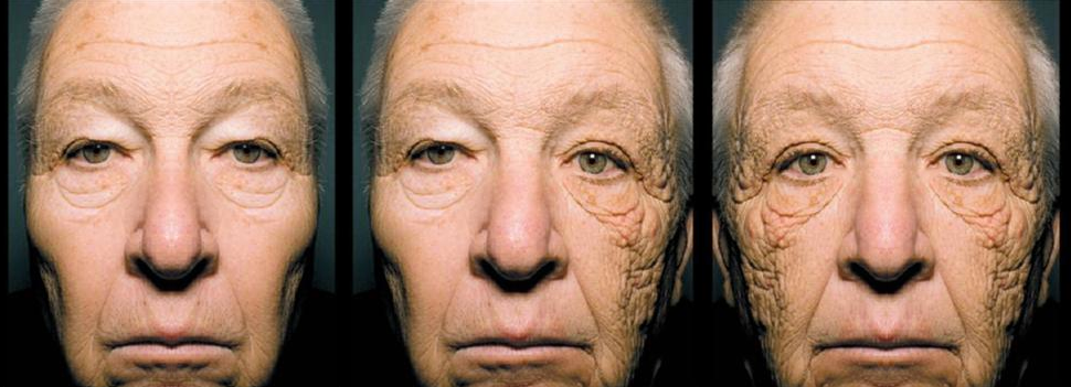
In the pictures above, you can see Mr. McElligott’s original picture in the middle with an altered image on either side. I transposed one side of his face to that of the same side in an attempt to make the difference between the two sides of his face more apparent.

Here you can find an “up-close and personal” version of the center picture. (How cool are his eyes?!?)

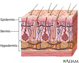
As you may already know, skin is made up of three major layers: the epidermis, dermis, and subcutis (also known as the subcutaneous layer or the hypodermis). As in the diagram below, the epidermis is on top, then the dermis, and lastly the hypodermis. Freckles and moles, as well as sunburns, appear in the epidermis. The dermis houses the structural elements in skin which keep it firm and elastic. This is the section of skin where wrinkles form.
Sunlight is known to negatively affect skin. This is because UV radiation (part of sunlight) can suppress the immune system, damage DNA, and alter skin texture, blood vessels and skin pigment. While conditions like thin skin and easy bruising are usually associated with old age, they are often lasting effects from UV damage as the majority of skin damage comes from the sun and not from Father Time.

The Sun emits three types of ultraviolet radiation: UVA, UVB, and UVC, with wavelengths of ~315 to 400 nm,  ~290 to 315 nm, and ~100 to 290 nm, respectively. UVC radiation is absorbed by the ozone layer and doesn’t reach ground level. UVB radiation makes its way through the ozone layer and is the type of UV that hits the epidermal layer of skin and causes sunburns or tans; it is also known to cause skin cancer. Unlike UVA radiation which is relatively constant all year long, UVB radiation is stronger during the summer months. UVA is unique in its ability to pass through glass. Thinking back to Mr. McElligott, UVA radiation’s ability to penetrate glass led him to have a build up of UVA damage of the course of his career. Even on days when he drove with his window up, he would have been protected from UVB rays, perhaps not getting a sunburn or tan, but would have still received the negative effects of UVA. UVA rays cause damage to the dermis, destroying elasticity and potentially causing mutations in DNA. Out of all of the UV radiation that reaches us from the Sun, there is over 20 times more UVA than UVB.

Harm to the skin caused by the Sun’s UV radiation is often referred to as photoaging, or dermatoheliosis. Photoaging is caused when UVA and UVB rays enter the skin, negatively affecting two of skin’s structural proteins, elastin and collagen. Radiation damages elastin and causes a build-up of altered metalloproteinase enzyme. Normally, metalloproteinases (also known as collagenases) help reverse skin damage by creating new collagen. Unfortunately, the metalloproteinase from the damaged elastin does the exact opposite— it breaks down collagen, leaving the skin with a broken collagen structure and wrinkles. (For a study on the effects of metalloproteinase,  check out this article.)

Exposure to UV radiation in sunlight, in addition to air pollution and smoking/second-hand smoke, can also subject the skin to free radicals. Free radicals are molecules (or atoms) with a single, unpaired electron. Because a free radical lacks the second electron, it is unstable and thus seeks out another electron. When it reaches another molecule, the free radical takes an electron, completing its electron pair while leaving the second molecule with an unpaired electron (a new radical). This snowball effect continues throughout the skin, activating altered metalloproteinase and causing damage to collagen.
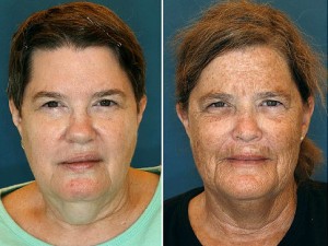
Jeanne (left) and Susan (right), identical twin sisters, serve as a great example of how lifestyle choices can determine a person's skin aging. As children, the two women had an uncanny resemblance, but now it's easy to see that Susan's decision to sunbathe and smoke led her skin to age more rapidly. (Department of Plastic Surgery, University Hospitals Case Medical Center)

Well, I hope you enjoyed learning more about wrinkles and UV radiation! Remember, prevention is key! Make sure to slather up in some good ol’ broad spectrum SPF 70 next time you go shoot some hoops (or do whatever it is that you do) and enjoy this fine video about why we need the Sun (even though it hurts us sometimes):


Sources:

Heather Brannon, MD (About.com)

Jennifer R.S. Gordon, MD and Joaquin C. Brieava, MD (The New England Journal of Medicine)

De Gruijl, Frank R. “Photocarcinogenesis: UVA vs. UVB Radiation.” Skin Pharmacology and Physiology 15.5 (2002): 316-20.  <http://www.ncbi.nlm.nih.gov/pubmed/10907526>.

Leave a comment

Your journey starts here! Let’s talk!

ThemeRex © 2026. All rights reserved.
  Timothy Durham was convicted of rape when a test showed his DNA genotype matched the DNA recovered from the crime scene. Eye witnesses testified that Durham was in a different state at the time of the incident, but he wasn’t exonerated from his 3000 year sentence until follow up DNA analysis showed that the preliminary forensic analysis used in the trial was misinterpreted (Thomas et al.). DNA testing serves an invaluable part in our criminal justice system, but it comes with complications and bias that can lead to wrongful convictions and misinformed juries.  

Background:

Forensic genetics aims to identify the origin of a biological sample. It can be traced back to Karl Landsteiner who discovered human ABO blood groups. With blood groups, suspects could be eliminated based on their blood type. For example, if a suspect had type A blood and there was type B blood left at the scene by the perpetrator, the investigators could rule out the suspect. Unfortunately, the reverse didn’t work. If the suspect and perpetrator both had the same blood type, it can’t be concluded that the suspect was guilty. Blood identifiers evolved into 8 distinct categories which improved the ability to eliminate suspects, but could only conclude that (if the system matched) it might be the perpetrator or this person might be related to the perpetrator (Jobling and Gill). The field evolved in the 1980s with the discovery of Minisatellites, lines of repetitive DNA that don’t encode proteins. Minisatellite patterns in DNA create what we know today as the DNA fingerprint, and it has a much higher accuracy rate than previous methods (Augustyn et al.). Around the same time, a breakthrough for victims of sexual assault was discovered. In the case of assault, bodily fluids of both the victim and the assaulter can get mixed. A process called differential lysis was created which could selectively increase the concentration of sperm in a fluid sample thereby allowing for accurate DNA testing (Jobling and Gill). Forensics evolved to use single-locus probe testing after several years because of its ease of interpretation. It looked at specific minisatellites that had high variability between subjects. This was the first DNA-based method to be used in a criminal investigation. Two murder homicides from several years apart were connected using DNA evidence. The evidence was then used to exonerate an innocent man, and the guilty party was found when he tried to evade mandatory DNA testing for all men in the small town. His guilt was then reinforced when the DNA profiles matched, and he gave a confession (Jobling and Gill). Finding DNA at a crime scene doesn’t guarantee that you’ll find a murderer. There are issues with non-pure samples and limited samples that make DNA work difficult, and accreditation of facilities and new techniques and take time (Jobling and Gill).  

Subjectivity of DNA Interpretation:

In a 2009 publication, the National Academy of Science discusses the possibility for subjective influence in lab based forensics. The interpretation bias happens in complex cases where, for example, there could be a mixture of DNA. A study published in 2011 looked specifically at this issue. A gang rape happened in Georgia, and the mixture in DNA from the case was used to prosecute one suspect after another suspect took a plea bargain to testify against him. With DNA mixtures, there are three possible conclusions: suspect excluded, suspect cannot be excluded, and inconclusive. If the suspect is excluded, it means that there’s little to no possibility that his DNA is part of the sample. If the suspect cannot be excluded, it means that there’s a possibility that his DNA is part of the sample. In the trial, the DNA examiners who knew the context of the case concluded that the suspect being prosecuted could not be excluded. The 2011 study gave the DNA evidence from this case and the DNA profiles of the suspects to 17 forensic examiners. These examiners had no context of the case, and they all worked independently. Of the 17, only 1 came to the same conclusion (suspect cannot be excluded) as the examiners who knew the context of the case. Twelve of the 17 concluded that the suspect who was prosecuted and convicted had no relation to the DNA found in the sample. Not only does this show that subjectivity occurs due to the context of the case, but the 17 examiners came up with different findings based on their past experiences meaning that not all DNA analysis is objective. Some organizations like the Scientific Working Group on DNA Analysis Methods say that quantitative statistical evidence should be presented with every categorical conclusion, but the US doesn’t require this (Dror and Hampikian).

False Positives:

There are two categories that cause wrongful identification in a DNA report: coincidental matches and false positives. Coincidental matches are DNA matches between two unrelated people. The probability of these matches is about 1 to 500,000 for white individuals and about 1 to 350,000 for black individuals. This small probability of coincidental matches makes people feel confident in DNA evidence. False positives, on the other hand, are matches between DNA and test DNA that result from issues in the forensics lab. There have been multiple cases of forensics labs accidentally switching victim and suspected rapist DNA which lead to convictions that were later overturned when the lab realized the error. There are also cases of misreading or misunderstanding a sample. In a case from 1993, a man was convicted of rape because someone interpreted a mixed sample of victim and perpetrator DNA as an individual sample. The conviction was again overturned. To prevent false convictions, retesting DNA is allowed, but it’s not an efficient solution. Many people are pushing for statistical information on the probability of false positives, so that the jurors can make accurate judgements based on DNA evidence (Thomas et al.).  

Conclusion:

DNA analysis is a useful tool for the criminal justice system, but subjectivity can be an issue in forensic testing. To combat wrongful convictions, there are organizations like The Innocence Project who use DNA testing to free the innocent, but reformed forensic testing standards should be put in place to prevent subjectivity and to ensure juries are correctly informed during trials. https://www.innocenceproject.org/    

Citation:

Augustyn , Adam, et al. “DNA Finerprinting.” Encyclopedia Britannica , 26 Apr. 2018, www.britannica.com/science/DNA-fingerprinting#ref153186. Dror, Itiel E., and Greg Hampikian. “Subjectivity and Bias in Forensic DNA Mixture Interpretation.” Science & Justice, vol. 51, no. 4, 2011, pp. 204–208., doi:10.1016/j.scijus.2011.08.004. Jobling, Mark A., and Peter Gill. “Encoded Evidence: DNA in Forensic Analysis.” Nature Reviews: Genetics, vol. 5, no. 10, Oct. 2004, pp. 739–751., doi:10.1038/nrg1455. Thompson, William C, et al. “How the Probability of a False Positive Affects the Value of DNA Evidence.” Journal of Forensic Sciences, vol. 48, no. 1, Jan. 2003, pp. 1–8., doi:10.1520/jfs2001171.