Day: April 24, 2026

  • What if humans weren’t meant to be here… like at all

    What if humans weren’t meant to be here… like at all

    What if humans weren’t meant to be here… like at all

    What if the first human to ever think for themselves didn’t ask how to survive… but instead asked why they existed at all?

    According to scientists working in these disciplines, humans have an inherent nature which pushes them towards finding explanations and developing patterns in their environment especially when faced with the unknown. Long before science, early cultures relied on rational stories for understanding their environments.

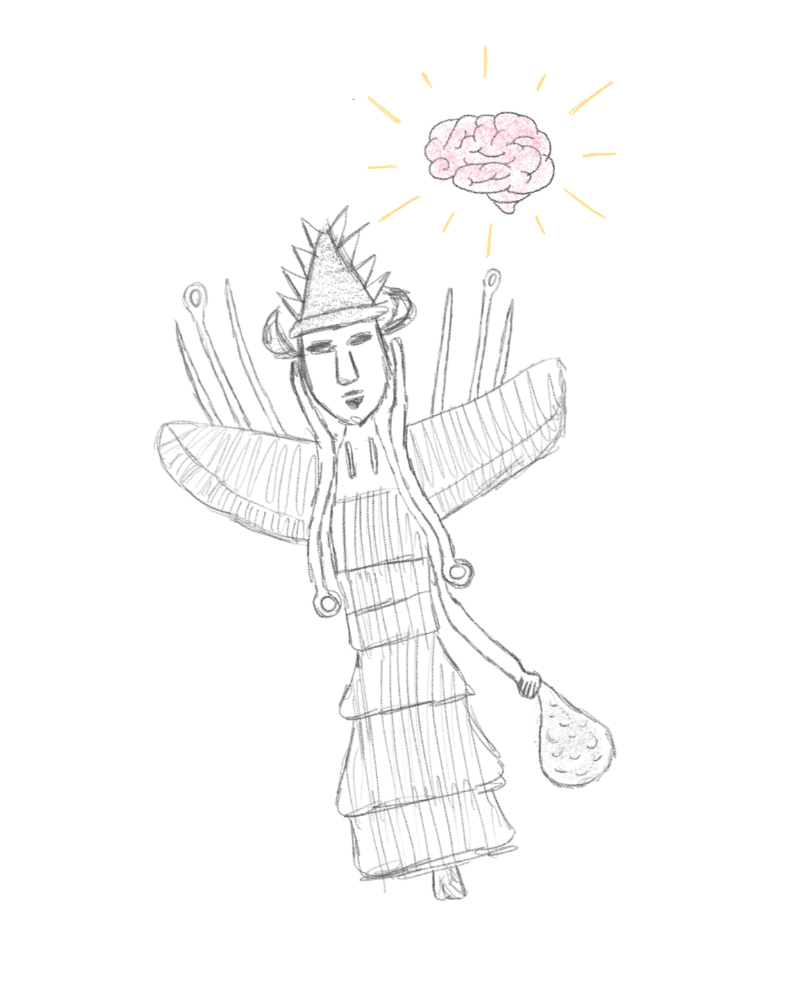
    Another such example is the one found in ancient Mesopotamian mythology. There are tales about otherworldly beings known as the Anunnaki who existed within this mythology. Not only did these beings exist within the stories, but they were crucial to comprehending the nature of human existence on Earth. Some Annunaki were associated with the sky, others to the underworld, and they were believed to decide human fate. As per the information provided by Encyclopaedia Britannica, Anunnaki refers to the offspring of god Anu who was a component of a cosmology that explained a wide range of occurrences in nature and human experience (Encyclopaedia Britannica, n.d.). Thus, it is necessary to note that, initially, knowledge about extraterrestrial beings did not exist.

    But something happened later on.

    The story didn’t stay the same.

    In the context of reinterpretation of the old myths in a modern setting, the whole story behind them got an entirely new meaning. All of a sudden, Annunaki was not just an ancient god anymore but an extraterrestrial being that was part of an incredibly advanced civilization experiencing some difficulties. To be more precise, the atmosphere became extremely polluted, which led to death. They turned to Earth because of one key resource: gold. Earth, rich in natural elements, became their solution.

    But there was a problem.

    Mining gold would take time. A lot of time.

    In order not to perform these duties by themselves, they created an instrument that would be able to work on their behalf.

    Humans.

    The Anunnaki merged their genes with those of hominids in order to create a species that would be capable of performing these tasks for them. At first, it worked. Humans followed the orders that were given. They mined, they built, they functioned almost like tools. But then something changed.

    One of the most compelling parts of this theory is the idea that one of these beings, often linked to Anu–the son of the Annunaki king–had a child with a human woman. This child was different. More aware, more knowledgeable. Picture yourself as the very first human asking, “Why am I doing this… and for whom?” That question alone challenges everything. Humans began to think deeply. They became emotional, unpredictable… aware. 

    But that’s the important thing: this entire tale of aliens and gold mining never appears in any Mesopotamian texts from antiquity. It is rather a theory that doesn’t originate from any scholarship. The original myths never described humans as engineered workers or the Anunnaki as extraterrestrial beings. That version of the story developed much later.

    So why does it still exist?

    Based on scientific analysis, the tendency among people to come up with origin stories is innate. According to cognitive science and anthropology, there is an inherent tendency among humans to develop structured explanations, often involving supernatural elements, since these allow humans to make sense of complexity and provide meaning to their existence (Boyer, 2001). Put simply, the Anunnaki story does not arise from thin air but from an instinctive drive in humans to make sense of their surroundings.

    But when science comes into play, the answer becomes obvious—and totally opposite.

    There are no scientific proofs to support the notion that extraterrestrials created humans. Instead, human evolution is explained through natural selection. Primitive forms of hominids, which refer to primitive people, slowly evolved from ancient human-like species into modern-day humans. It is clear that according to the evidence provided by fossil records and genetic researches, Homo sapiens are believed to have originated from various animals that existed in the past due to the effects of natural selection (Stringer, 2016). According to scientists, natural selection is a process through which particular traits allow for survival and reproduction, which leads to improved behavioral patterns and neurological development. 

    This process was not planned. It was not designed.

    It was shaped by the environment.

    Even the idea of gold—so central to the Anunnaki theory—has a scientific explanation. The presence of gold on Earth was not intentional. Rather, gold was formed in space due to some calamities like supernovae or neutron stars billions of years back. In the following period, geologic phenomena such as volcanic eruptions led to the presence of gold in Earth’s crust. In other words, the existence of gold on Earth was not because any being had come down to earth for mining gold; it was the creation of the universe that made it possible.

    What does this mean for us then?

    The myth concerning the Anunnaki can be said to be in between a myth and scientific theory. Ancient myths explained many things for ancient people. But modern theories reinterpret ancient myths in the context of science. And science explains everything, though perhaps not very mystically.

    But perhaps that is the whole idea.

    For it is not only about determining the truth regarding the existence of the Anunnaki.

    It’s about something much deeper in the human mind.

    Humans have always been in pursuit of some sort of meaning. There is a constant inquiry into the reason for our existence, our difference from other animals, and our purpose. These ideas show how far we are willing to go to explain our existence.

    So maybe the real question isn’t “Were the Anunnaki real? Did they really create humans?”

    Maybe the real question is…

    “Why do we keep wanting a story where something created us—on purpose?”

    References (APA 7th Edition)

    Boyer, P. (2001). Religion explained: The evolutionary origins of religious thought. Basic Books. https://sackett.net/BoyerReligionExplained.pdf

    Encyclopaedia Britannica. (n.d.). Anunnaki. https://www.britannica.com/topic/Anunnaki

    Stringer, C. (2016). The origin and evolution of Homo sapiens. Philosophical Transactions of the Royal Society B, 371(1698). https://royalsocietypublishing.org/rstb/article/371/1698/20150237/22900/The-origin-and-evolution-of-Homo-sapiensThe-origin

     

  • Summer Nostalgia: Why are Scientists Studying how Fireflies Glow?

    Summer Nostalgia: Why are Scientists Studying how Fireflies Glow?

    Introduction:

    You step outside on a warm summer night to see a dark sea with little, intermittent flashes of light. Catching fireflies is a nostalgic childhood memory for people all across the globe. But what allows these bugs to produce this soft glow? 

    Fireflies produce their light through bioluminescence, an efficient light production process resulting from a chemical reaction. While the light comes naturally from fireflies, scientists are trying to understand how to replicate this process for medical research applications with the same ease as fireflies.

    How does this chemical reaction work?

    While the chemical reaction itself is relatively straightforward, fireflies have to carefully control when to light up. This bioluminescent process takes place in a special light producing organ in the abdomen. Within this organ, adenosine triphosphate (ATP), which stores energy in living cells, combines with oxygen, calcium, and luciferin with luciferase, the fireflies emit light (Branham, 2005).

    Fireflies can control the flow of oxygen into the photocytes, which is part of the light-producing organ. When oxygen enters the photocytes, it reacts with the luciferin in the presence of luciferase, an enzyme, and ATP and produces light (Martin, n.d.). When oxygen is allowed to flow into the photocytes, light is emitted.

    Recent studies suggest that fireflies evolved to have biochemical pathways for the production of luciferin and the regulation of the reaction (Zhang et al., 2020). This allows for the fireflies’ bioluminescent system to be highly effective as the energy dissipated from this reaction is emitted as light instead of heat (Zhang et al., 2020). This efficiency has proven to be difficult to replicate without these adaptations, which is why scientists are trying to recreate the process in a laboratory.

    How does this Chemical Reaction Work in the Lab?

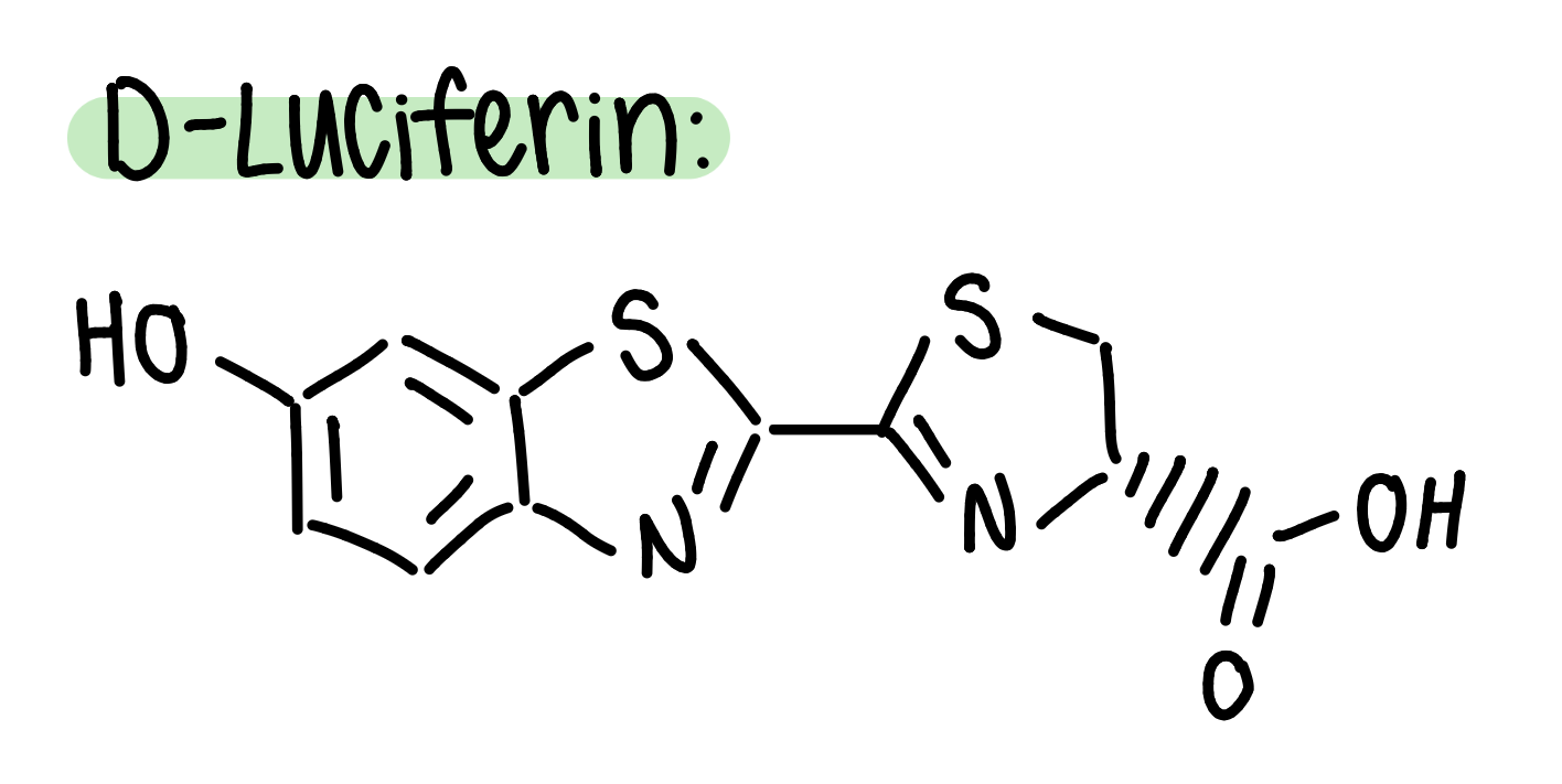
    Researchers at Nagoya University in Japan have created a six-step synthesis for D-Luciferin, which is the specific stereoisomer, or orientation, of luciferin that binds with luciferase to produce light using p-benzoquinone, L-cysteine methyl ester, and D-cysteine (Kato et al., 2025). 

    This synthesis is designed to replicate the natural production of luciferin with greater efficiency and lower costs. The synthesis is done in a one-pot reaction to streamline the process and make it applicable on a larger scale (Kato et al., 2025). While this synthesis has successfully yielded D-luciferin, creating an artificial version of the full biological system of fireflies, especially with the control of oxygen flow, remains a challenge. 

    The efforts of the researchers at Nagoya University show the complexity of replicating a naturally optimized biochemical process. But why are scientists investing so much into recreating firefly bioluminescence? 

    Why are Scientists Studying this Process?

    Scientists have investigated the mechanism of the bioluminescent process of fireflies due to the emission’s application in medical imaging, including cancer research as a way to locate tumors (Martin, n.d.). However, the cost of producing the same reaction that fireflies do every night, every summer for free is quite expensive, driving the motivation for scientists to create the most efficient, cost and time-wise, synthesis for D-Luciferin (Kato et al., 2025). 

    Conclusion

    What appears to be a piece of summer nostalgia is actually a highly optimized biochemical process. By controlling the reaction between luciferin, luciferase, ATP, and oxygen, fireflies are able to make a chemical reaction a mesmerizing scene. 

    Scientists are studying the process of firefly light production to make the synthesis of D-luciferin more accessible for research and medical applications. The same soft glow that lights  up a warm summer night is now contributing to science and medicine, showing how even the smallest of animals and insects can lead to scientific progress. 

    References:

    Branham, M. (2005, September 5). How and why do fireflies light up? Scientific American. Retrieved April 17, 2026, from https://www.scientificamerican.com/article/how-and-why-do-fireflies/

    Kato, M., Tsuchihashi, K., Kanie, S., Oba, Y., & Nishikawa, T. (2025, December 25). A practical, biomimetic, one-pot synthesis of firefly luciferin. Nature Science Reports, 14(1). https://doi.org/10.1038/s41598-024-82996-2

    Martin, K. (n.d.). Everything About Luciferin and Luciferase. GOLDBIO. Retrieved April 17, 2026, from https://www.goldbio.com/blogs/articles/everything-about-luciferin-luciferase?srsltid=AfmBOoo5RwdWL6Aa1C7Lu7UK2e9LLM0KLTmjIYMHJmoU7DI5741Ww0Qa

    Zhang, R., He, J., Dong, Z., Liu, G., Yin, Y., Zhang, X., Li, Q., Ren, Y., Yang, Y., Liu, W., Chen, X., Xia, W., Duan, K., Hao, F., Lin, Z., Yang, J., Chang, Z., Zhao, R., Wan, W., … Li, X. (2020, September 28). Genomic and experimental data provide new insights into luciferin biosynthesis and bioluminescence evolution in fireflies. Nature Science Reports, 10(1). https://doi.org/10.1038/s41598-020-72900-z

  • How Pymol Lets You Visualize DNA in 3D

    How Pymol Lets You Visualize DNA in 3D

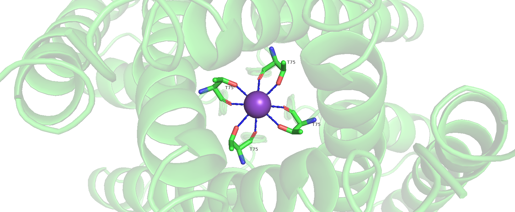
    Have you ever wondered how scientists look at proteins or DNA? Here’s the secret- they don’t actually look at physical molecules all that often! Frequently, they use softwares like PyMOL. PyMOL is a widely used molecular visualization program that converts structural data into detailed 3D models, allowing scientists to rotate, zoom, and explore molecules interactively. It was originally developed by Warren Lyford DeLano and has become a standard tool in structural biology and chemistry (DeLano, 2002).

    PyMOL works by reading files that contain the exact positions of atoms in three-dimensional space, often obtained from experimental techniques like X-ray crystallography and stored in databases such as the Protein Data Bank. Using this coordinate data, the program reconstructs molecules and displays them in different visual styles, including sticks, spheres, and ribbon diagrams that highlight protein folding patterns. These visualization approaches are essential for understanding how molecular structure relates to function and are widely used in both research and education (Sehnal et al., 2021).

    What makes PyMOL especially powerful is how interactive and customizable it is. Scientists can highlight specific regions of a molecule, measure distances between atoms, and even model how molecules might interact with each other- something that is especially important in drug design and biochemistry. Because it translates complex data into intuitive visuals, PyMOL helps bridge the gap between abstract molecular concepts and real-world understanding, making it a valuable tool not just for scientists, but also for students learning about the molecular world.

    References:

    DeLano, W. L. (2002). PyMOL: An open-source molecular graphics tool. San Carlos, CA: DeLano Scientific. Available at http://www.pymol.org

    Sehnal, D., Bittrich, S., Deshpande, M., Svobodová Vařeková, R., Berka, K., Bazgier, V., Velankar, S., Burley, S. K., Koča, J., & Rose, A. S. (2021). Mol* Viewer: Modern web app for 3D visualization and analysis of large biomolecular structures. Nucleic Acids Research, 49(W1), W431–W437. https://doi.org/10.1093/nar/gkab314

    Berman, H. M., Westbrook, J., Feng, Z., Gilliland, G., Bhat, T. N., Weissig, H., Shindyalov, I. N., & Bourne, P. E. (2000). The Protein Data Bank. Nucleic Acids Research, 28(1), 235–242. https://doi.org/10.1093/nar/28.1.235

  • How to make glow in the dark cells

    How to make glow in the dark cells

    While these cells don’t technically glow in the dark, scientists have found ways to make cells fluorescent which means they glow when exposed to radiation such as UV light. When a cell has specific ions or proteins fluorescent proteins bind to these small molecules and make the cell appear as if it’s glowing. 

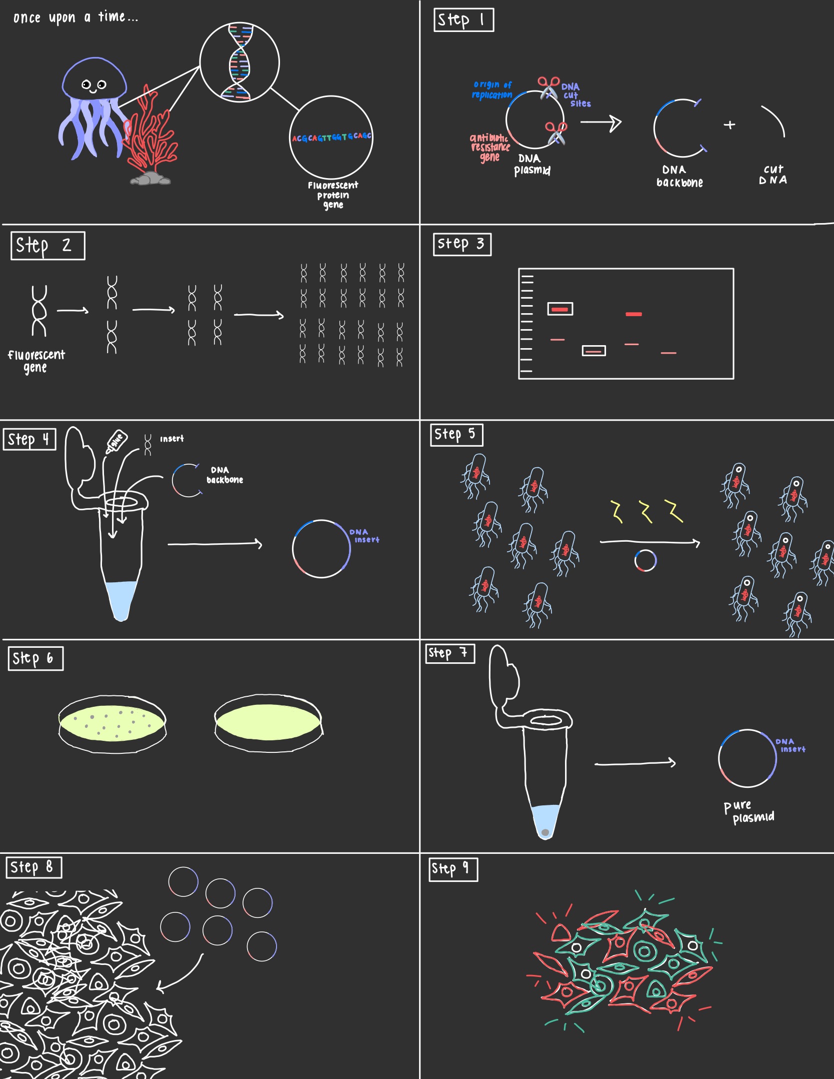
    These special proteins were first discovered in animals like jellyfish and plants like coral. Scientists were able to isolate the genes that were responsible for producing the fluorescent proteins and using a technique called molecular cloning can insert the DNA into our cells of choice. 

    Step 1: The first step is to choose a plasmid, a circular piece of DNA found in bacteria. The plasmid chosen must have an origin of replication where DNA replication begins, a gene for antibiotic resistance, and DNA cut sites. Restriction enzymes are proteins that act as molecular scissors and cut the plasmid at specific cut sites. This turns the circular plasmid DNA into a linear piece of DNA. 

    Step 2: This isn’t always necessary, but the fluorescent gene can be amplified using a technique known as polymerase chain reaction. In a PCR the DNA undergoes cycles of denaturation, annealing, and elongation where the DNA is first turned into single stranded DNA, then the primer tells the DNA where to start extending, and then finally the DNA is extended. At the end of each cycle the amount of DNA is doubled leading to millions of copies of DNA within an hour. 

    Step 3: By this point, we have our amplified PCR DNA and our plasmid DNA that has been cut to produce the backbone and pieces of cut DNA. In order to separate the backbone plasmid from the cut piece we will use gel electrophoresis. A gel electrophoresis separates DNA by size by passing an electric current through a gel containing DNA. Because DNA is negatively charged it will migrate towards the positively charged terminal of the gel with the smaller pieces traveling further than the larger pieces. Because our backbone is larger than the cut DNA, when the pieces separate we can cut out the band closer to the top, and purify it to get rid of the gel leaving just the DNA. 

    Step 4: It’s finally time to glue our DNA pieces together, and the key player is a protein known as DNA ligase. DNA ligase will be added to a tube with our DNA backbone and insert. Following a ligation reaction a plasmid will be re-formed that contains our original backbone with the fluorescent gene inserted in. 

    Step 5 and Step 6: The next two steps are crucial for ensuring that the final product is pure plasmid. This is important because the DNA ligase can make mistakes and may glue backbones together, fail to add in the insert, etc. In step 5 the plasmid is inserted into bacterial cells, usually e-coli cells through a heat shock. Once the bacteria take up the plasmid they are plated onto agar plates containing antibiotics and allowed to grow. Remember the three things every plasmid needs to include (hint: it’s an antibiotic resistance gene)? This is because when the cells are plated only those that have a fully formed plasmid will be able to grow. 

    Step 7: Now we can select a bacterial colony from our plate and using a process known as miniprep isolate the plasmid DNA from these cells. 

    Step 8: The plasmid DNA can then be introduced into your cells of choice. There are many techniques to accomplish this but a common one is called calcium phosphate transfection. 

    Step 9: TA-DA! Your cells should now glow under the right conditions! 

    Sources:

    Ashwini, M., Murugan, S.B., Balamurugan, S. et al. Advances in molecular cloning. Mol Biol 50, 1–6 (2016). https://doi.org/10.1134/S0026893316010131

    Tsien RY. The green fluorescent protein. Annu Rev Biochem. 1998;67:509-44. doi: 10.1146/annurev.biochem.67.1.509. PMID: 9759496.
    cover image created using canva
  • Microscopic Masterpiece

    Microscopic Masterpiece

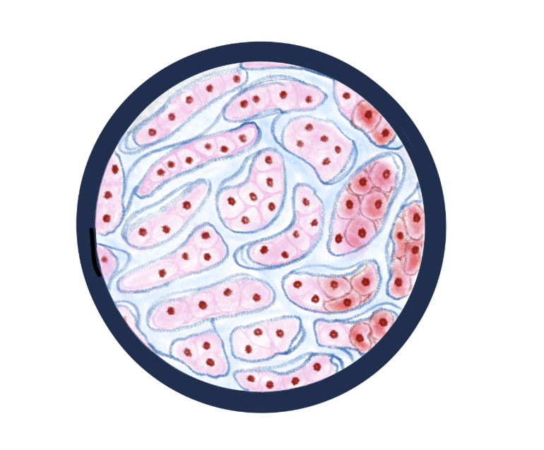
    This may be bad to admit, but I love a good TikTok scroll. It’s probably not the best use of my time, but I’ve come across some interesting things! New music artists to listen to, events around my city, and even some science accounts. A bit ago, I found a medical student who makes histological artwork and I was absolutely fascinated. Her name is Kait and her paintings are absolutely beautiful (@createbykait on TikTok if you are curious)!

    Of course then I had to do a deep dive about histology. What actually is it? What is she really painting? I assumed it was cells from the look of it, but that was the extent of my knowledge. Turns out there is a lot more to histology than just a pretty picture.

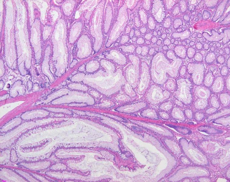
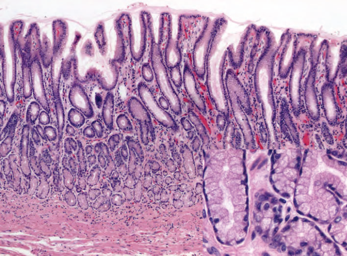
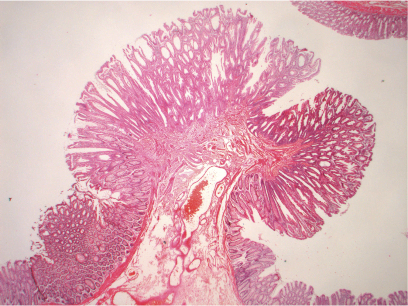
    Histology is the study of microscopic structures of tissues. It usually includes sectioning a thin slice of tissue, staining it with dyes that adhere to different types of structures, then observing the slice under a microscope (NCBI Bookshelf, 2023). Histology is also the foundation for pathology, which is the study of the effects of disease on the body. I had heard pathology as a medical specialty, but again, did not know what they did.

    Turns out, pathologists look at tissue samples from biopsies or autopsies to make diagnoses. I got to watch a brain biopsy earlier this year while shadowing a neurosurgical oncologist. These patients had gotten MRIs that showed a spot of something irregular, called a lesion. During the biopsy, the surgeons took a small sample of the lesion tissue to send to a pathologist to determine what it was, whether it was cancer, an aneurysm, diseased tissue, or even a parasite. The first sample they took is called the frozen section, which they would send to pathology immediately. The pathologists would quickly freeze the sample and examine it to make sure it was usable. The surgeons would have to stay scrubbed in and wait in the operating room until they got confirmation from the pathologist that the sample was good. If it wasn’t, the surgeons would go in and get another sample before closing the wound.

    Pathologists also examine autopsies, or tissue from dead organisms. This is often used when there is an investigation with an unclear cause of death. This led me to Marianne Hamel, a forensic pathologist, who recognized how paradoxically beautiful these histological images can be. So much so that she created a collection of histological images that is now on display at Rudger’s University (Hamel & Johnson, n.d.). She said in an interview that she hopes to educate people about her career field, but also allow people to appreciate the beauty of the human body (Chandler, 2023).

    Histology has evolved a lot since its beginnings. Hamel said photos used to be shot in black and white, making interpreting the images much more difficult. Now there is a plethora of different dyes used to make multicolored images. This offers so much more information for pathologists to gather, but also a higher level of difficulty. Learning to read [histological] slides, Hammel says, is like learning another language (Chandler, 2023).

    It’s funny how what seems like just a pretty picture is actually such an intricate science and can show how beautiful life can be.

    “Mixed histology polyp.” Image by Caoimhin74, via Wikimedia Commons, licensed under CC BY-SA 3.0.
    “Histology of normal antral mucosa” Image by Carrasco G, Corvalan AH, via Wikimedia Commons, licensed under CC BY-SA 3.0.
    “Mixed histology polyp” Image by Caoimhin74, via Wikimedia Commons, licensed under CC BY-SA 3.0.

    References

    National Center for Biotechnology Information. (2023). NCBI Bookshelf. U.S. National Library of Medicine. https://www.ncbi.nlm.nih.gov/books/NBK557663/

    Hamel, M., & Johnson, N. (n.d.). Death under glass. https://www.deathunderglass.com/

    Chandler, R. (2023, October 31). Post-mortem histology artwork brings death to life. Evident Scientific. https://evidentscientific.com/en/insights/post-mortem-histology-artwork

  • Matcha: More Than Just a Trend

    Matcha: More Than Just a Trend

    After going viral on social media in the early to mid 2020s, matcha – a traditional Japanese green tea drink – now has a ubiquitous presence in many cafes and even bakeries. Popular for its aesthetic green color and unique earthy taste, matcha’s global market has exploded over a short period of time and was estimated to be worth 5.07 billion USD in 2025 (Grand View Research, 2023). Along with its popularity, there has been growing interest in its potential health benefits. 

    In fact, recent research has suggested that matcha may actually have several health benefits – such as increasing cognitive performance, decreasing stress and anxiety, and even reducing inflammation. 

    What is matcha exactly?

    Matcha is the powdered form of Japanese green tea and has been mostly consumed in traditional settings as well as incorporated into many food items. Originating from the Camellia sinensis plant, it has been traditionally cultivated by shielding the tea bushes from the sun using bamboo mats and steamed afterwards to prevent oxidation (Sokary et al., 2023). This process allows for higher production of amino acids and compounds like chlorophyll, which gives matcha its dark and vibrant green color as well as umami flavor (Sokary et al., 2023). 

    In addition, matcha’s chemical composition consists of polyphenols (aka very powerful antioxidants), catechins, caffeine, and phenolic acids. It also contains theanine, which is a non-protein amino acid that is known to have anti-stress effects (Baba et al., 2021). Notably, it is believed that the combination of theanine and caffeine in matcha, which is not found in coffee, helps improve attention (Baba et al., 2021). 

    Benefits of Matcha

    A recent study researched the effects of matcha on executive functioning such as attention (Baba et al., 2021). The participants were randomly assigned to three groups, which were placebo, caffeine, and matcha (Baba et al., 2021). They consumed 9 pills – either placebo, caffeine, or matcha – for 12 weeks every morning and afterwards, were tested on several skills (i.e., memory, facial expression recognition, attention, etc.) using a set of cognitive tests (Baba et al., 2021). The results from this experiment found that the matcha group demonstrated significantly lower reaction times and increased perception of emotion (Baba et al., 2021). In addition, the same group conducted another experiment to observe the effects of matcha under mildly stressful conditions. They found that continuous consumption of matcha helped improve performance under stress (Baba et al., 2021).

    Another study conducted an experiment that examined matcha’s effect in mice exposed to air pollution. All mice were exposed to particulate matter before the experiment. The experimental group of mice were given 20 or 40 mg/kg of matcha, which was based on body weight (Jong Min Kim et al., 2021). Afterwards, it was found that matcha helped to alleviate inflammation (Jong Min Kim et al., 2021). The researchers also found that the experimental (matcha) group was protected from antioxidant deficits in several tissues of the body (Jong Min Kim et al., 2021).

    Conclusion

    Beyond its trendiness and aesthetic, matcha can be described as a delicious concoction of various nutrients, ranging from antioxidants to amino acids that help prevent inflammation and even reduce stress. So the next time you find yourself stressed or need to sharpen your attention skills, head on to a nearby cafe and try a matcha!

    References

    Baba, Y., Inagaki, S., Nakagawa, S., Kobayashi, M., Kaneko, T., & Takihara, T. (2021). Effects of Daily Matcha and Caffeine Intake on Mild Acute Psychological Stress-Related Cognitive Function in Middle-Aged and Older Adults: A Randomized Placebo-Controlled Study. Nutrients, 13(5), 1700. https://doi.org/10.3390/nu13051700

    Grand View Research. (2023). Matcha Market Size, Share | Global Industry Trends Report, 2018 – 2025. Www.grandviewresearch.com. https://www.grandviewresearch.com/industry-analysis/matcha-market

    Jong Min Kim, Jin Yong Kang, Seon Kyeong Park, Jong Hyun Moon, Min Ji Kim, Hyo Lim Lee, Hye Rin Jeong, Jong Cheol Kim, & Ho Jin Heo. (2021). Powdered Green Tea (Matcha) Attenuates the Cognitive Dysfunction via the Regulation of Systemic Inflammation in Chronic PM2.5-Exposed BALB/c Mice. Antioxidants, 10(12), 1932–1932. https://doi.org/10.3390/antiox10121932

    Kochman, J., Jakubczyk, K., Antoniewicz, J., Mruk, H., & Janda, K. (2020). Health Benefits and Chemical Composition of Matcha Green Tea: A Review. Molecules, 26(1), 85. https://doi.org/10.3390/molecules26010085

    Sokary, S., Al-Asmakh, M., Zakaria, Z., & Bawadi, H. (2023). The therapeutic potential of matcha tea: A critical review on human and animal studies. Current Research in Food Science, 6, 100396. https://doi.org/10.1016/j.crfs.2022.11.015

    Uchida, K., Meno, K., Tatsumi Korenaga, Liu, S., Suzuki, H., Baba, Y., Tagata, C., Araki, Y., Shuto Tsunemi, Aso, K., Inagaki, S., Nakagawa, S., Kobayashi, M., Tatsuyuki Kakuma, Asada, T., Ota, M., Takanobu Takihara, & Arai, T. (2024). Effect of matcha green tea on cognitive functions and sleep quality in older adults with cognitive decline: A randomized controlled study over 12 months. PLoS ONE, 19(8), e0309287–e0309287. https://doi.org/10.1371/journal.pone.0309287

  • Lament of a Musician’s Hearing

    Lament of a Musician’s Hearing

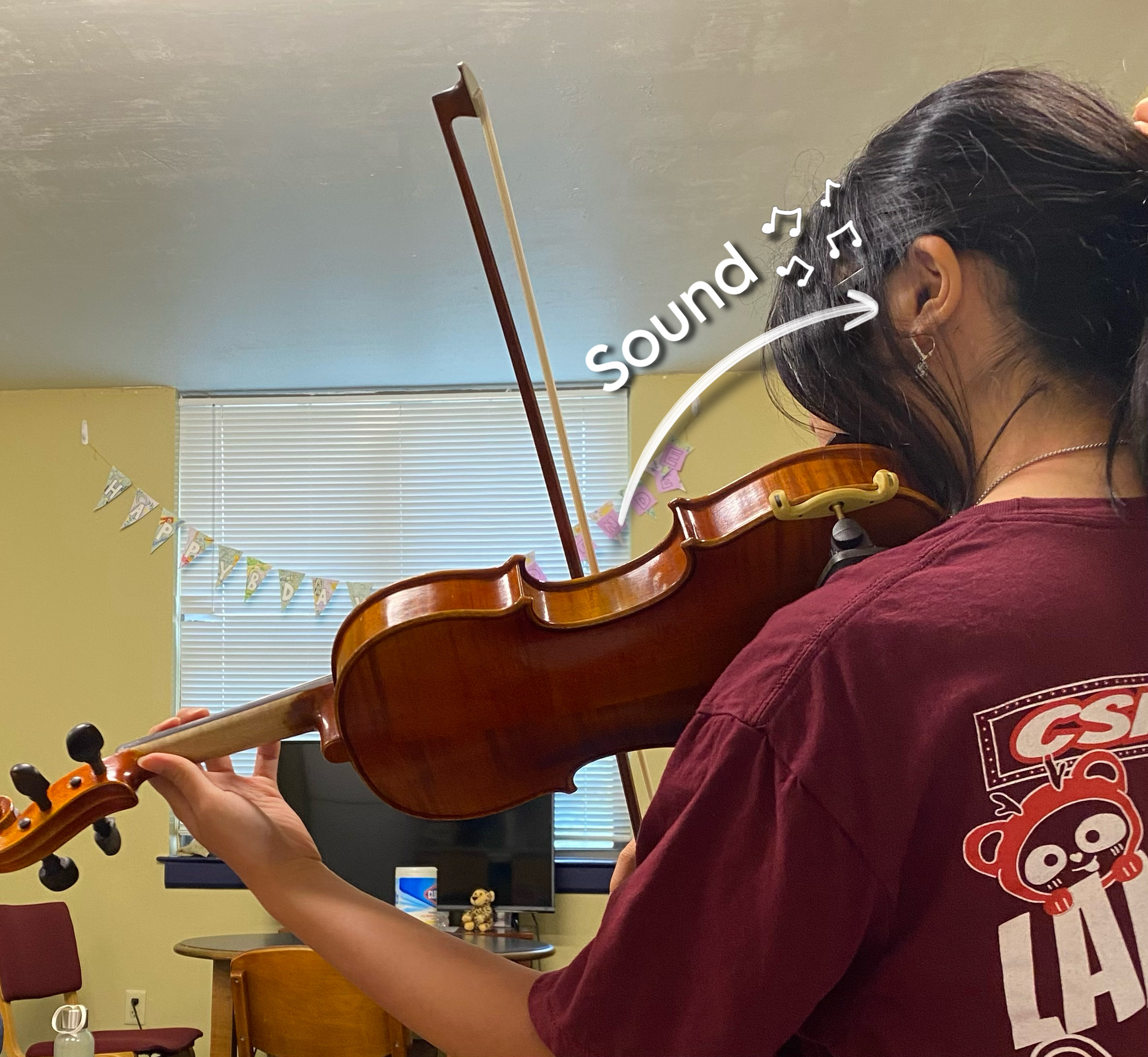
    Cover image was taken by the author and edited in Picsart.

    Sometimes, what is most important to us is what we take for granted. For a musician, that is their hearing, although most of us don’t take many steps to preserve it, often going to rehearsal or lessons without any form of hearing protection. For a school musician, this means going a few hours a week being exposed to loud noises without any way to block them, and for a professional, this duration is even longer. If we don’t want to lose our hearing to the piercing sound of a piccolo in our ears, it’s possible we might have to make some changes to the way we rehearse.

    Hearing Loss in Musicians Compared to Other Professions

    Some might question whether playing an instrument actually increases hearing loss or if there is a larger outside factor at play. To investigate this, a study was conducted comparing the hearing loss of people with a variety of different backgrounds and found that professional musicians were 3.51 times more likely to have noise induced hearing loss and 1.45 times more likely to have tinnitus (Schink et al., 2014), which suggests that the job that someone works in has a large impact on that person’s hearing. This makes sense considering the significant amount of time the average adult spends working. Musicians tend to get the short end of the stick here as a good portion of that time is spent listening to loud music, which oftentimes when playing is right in your ear.

    Source: taken by author and edited in Picsart.

    Reducing Ear Damage

    Since the consequences of continued exposure to loud noise can become severe over time, it is important to take steps in order to prevent excessive damage. First and foremost, musicians should be aware of the risks that long periods of practice can have on their hearing; they should be able to watch for warning signs and know what safe sound levels are (85 dB TWA (time-weighted average) for eight hours in one day, which is around the noise level of a food blender, heavy traffic, busy restaurant, or movie theater (How Loud Is 85 Decibels | What Does 85 DB Sound Like, 2021)). It is also beneficial to get an audiogram and have access to a sound-level meter to make sure you aren’t exceeding the safe sound levels (Chesky, 2008). Also, most students have some sort of hearing protection device or HPD (around 69% percent according to one study) that they can use during rehearsal to block some of the sound (Olson et al., 2016).

    Source: (Lowery, 2023)

    The Great Balance

    If the measures to be taken are seemingly so obvious and simple, why aren’t they more common? Even if the resources are readily available, it turns out that protecting one’s hearing still comes with its downsides. According to several music students, even though there are clear advantages to using things like HPDs, such as hearing preservation, reduction in noise exposure, and the ability to play longer without taking breaks, there are also significant disadvantages to consider like decreased auditory perception, issues with communication, less comfort, and many reported improved performance when not using HPDs (Olson et al., 2016). The goal of an ensemble is to make music that sounds good (well, and have fun, hopefully), so naturally, the musicians in an ensemble would not want to sacrifice their sound to protect their hearing, which is something they might not even be conscious of because damage happens gradually.

    So how do we balance the desire to play well with the desire not to go deaf before we get old? Perhaps this will be a question better answered in the future, maybe even by someone reading this article, and we’ll have a device that still lets us hear everything we need to without all of the damage. Maybe we’ll have a way to reverse hearing loss that’s as easy as drinking medicine to cure a cold. But for now, while maybe not during performances, we can just do what we can, however little it may seem, to protect one of our precious senses. We only have five, after all.

    References

    Chesky, K. (2008). Preventing Music-Induced Hearing Loss. Music Educators Journal, 94(3), 36–41. https://www.jstor.org/stable/4623689

    How Loud Is 85 Decibels | What Does 85 dB Sound Like. (2021, November 18). Decibel Meter App | Best Digital Sound Level Meter for Your Smartphone. https://decibelpro.app/blog/how-loud-is-85-decibels/

    Lowery, K. (2023). How to choose the best hearing protection [Online Image]. In audibel.com. https://www.audibel.com/preventative-care/the-best-hearing-protection/

    Olson, A. D., Gooding, L. F., Shikoh, F., & Graf, J. (2016). Hearing Health in College Instrumental Musicians and Prevention of Hearing Loss. Medical Problems of Performing Artists, 31(1), 29–36. JSTOR. https://www.jstor.org/stable/48714485

    Schink, T., Kreutz, G., Busch, V., Pigeot, I., & Ahrens, W. (2014). Incidence and relative risk of hearing disorders in professional musicians. Occupational and Environmental Medicine, 71(7), 472–476. JSTOR. https://www.jstor.org/stable/43869897