Treating U87 EGFP Glibolastoma Cells with Nickel Sulfate!

Part of the reward of being a Biomedical Engineer is the opportunity to explore fascinating and cutting-edge work that teaches me new skills and exposes me to unique challenges every semester. Throughout my journey, I’ve learned coding, worked with circuits, conducted detailed statistical analyses, and performed rigorous statistical tests. However, this semester, I had the chance to do something I had never experienced before. I worked with cancer cells. Yes, you read that right! I worked with cancer cells in a wet lab environment!

What is a wet lab, you ask? It’s a controlled environment where you work with potentially hazardous cell cultures and substances while following strict safety protocols, such as wearing gloves, goggles, lab coats, and disinfecting surfaces with ethanol to avoid contamination. Mistakes can introduce bacteria or other pathogens that could destroy your cultures, making careful adherence to procedure critical. Despite the challenges, working in a wet lab is an incredibly rewarding experience that not everyone gets to have. To share this unique experience, I want to provide some insight into what a typical experiment looks like in a wet lab, so those who haven’t had the opportunity can get a feel for it. Trust me, you’ll find it fascinating!

One of the experiments I conducted this semester involved treating Glioblastoma (U87-EGFP) cells with varying concentrations of Nickel Sulfate (NiSO₄) to assess its effects on cell viability. Intuitively, I hypothesized that increasing concentrations of Nickel Sulfate would be progressively toxic to these cells. Sure enough, that’s exactly what happened. The coolest part? I didn’t even need to run complex calculations to see the effects! By observing the cells under a microscope, I could identify the toxicity based on changes in cell morphology and network structure. Let me explain how this works.

When cancer cells are healthy, they exhibit a spherical shape and form densely interconnected networks, similar to what we see in normal, healthy cells. However, as cells are damaged, these networks weaken, and their shapes become less defined and less spherical. These morphological changes occur because interactions between the cells’ mitochondria and cytoskeleton are disrupted. Observing these changes during the experiment allowed me to visually interpret the effects of Nickel Sulfate on the cells. Below, I’ve described and analyzed the experiment’s results to demonstrate how the process unfolded in real time.
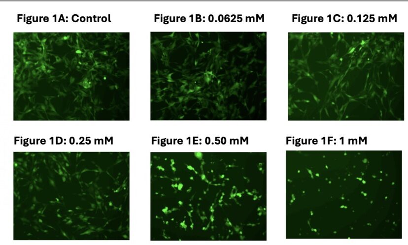
Figure 1A. Appearance of U87 – EGFP cells, after addition of 0 mM of NiSO4. Cells appear to be healthy, as they are exhibiting a bright color, are elongated in shape, and are forming an interconnected network like structure.

Figure 1B. Appearance of U87 – EGFP cells, after addition of 0.0625 mM of NiSO4. Cells still appear to behealthy, but they are slightly less elongated, and are slightly less bright in color, as compared to when 0 mM ofNiSO4 was added. The cells do still appear to be interconnected, though.

Figure 1C. Appearance of U87 – EGFP cells, after addition of 0.125 mM of NiSO4. Cells appear to be less healthy, and are significantly less elongated and bright, compared to when 0 or 0.0625 mM of NiSO4 wasadded. Cells also appear to be significantly less interconnected, as the network structure is weakening. The addition of NiSO4 at this concentration seems to be having a significant impact on the structure of NiSO4, indicating that the IC50 concentration is being approached.

Figure 1D. Appearance of U87 – EGFP cells, after addition of 0.25 mM of NiSO4. Cells appear to be significantly less healthy, and there is hardly any presence of elongation, compared to when 0, 0.0625, or even0.125 mM of NiSO4 was added. There also appears to be hardly any network structure left between the cells, as all interconnections are not visible. The cells are also significantly less bright, and are extremely dim. The significant structural changes that arise from the addition of this concentration of NiSO4 indicate that the IC50 concentration has been passed.

Figure 1E. Appearance of U87 – EGFP cells, after addition of 0.50 mM of NiSO4. Cells appear to be significantly less healthy, and do not have any visible interconnected network structure. Any slight presence of anetwork structure seen when 0.25 mM of NiSO4 was added also appears to be gone. Now, there appears to be no network structure.

Figure 1F. Appearance of U87 – EGFP cells, after addition of 1 mM of NiSO4. Cells appear to be significantly less healthy, as there is no elongation present. There is also no network structure left between the cells. No interconnections appear to be visible. The cells are in a completely opposite state to how they were, from a color perspective, when they were treated with no NiSO4. Given the permanent changes in structure and morphology of the cells, it is likely that the addition of 1 mM NiSO4 has the maximum possible impact on the cells. In other words, it is likely adding any more NiSO4 will have a marginal impact, if any.

Overall, observing these morphological changes was an eye-opening experience for me as it taught me to analyze cellular changes, in a relatively simple way, without relying on quantiative measurements or complicated formulas, something that I never knew that you could do as an Engineer! It also taught me that cancer is not as invincible as it is made out to be and it can indeed be quashed. This is something that I never knew until I did this experiment, and it gave me a lot of hope for the future of cancer treatment!

Reading this post, I hope you got inspiration to work in a Wet lab too one day as you see all the cool stuff that you can do/accomplish. Sometimes, you do not have to be a fancy scientist at the NIH or the CDC to make a difference. Occasionally, just the Georgia Tech student can accomplish some pretty cool stuff!


 

Your journey starts here! Let’s talk!

ThemeRex © 2026. All rights reserved.
  Timothy Durham was convicted of rape when a test showed his DNA genotype matched the DNA recovered from the crime scene. Eye witnesses testified that Durham was in a different state at the time of the incident, but he wasn’t exonerated from his 3000 year sentence until follow up DNA analysis showed that the preliminary forensic analysis used in the trial was misinterpreted (Thomas et al.). DNA testing serves an invaluable part in our criminal justice system, but it comes with complications and bias that can lead to wrongful convictions and misinformed juries.  

Background:

Forensic genetics aims to identify the origin of a biological sample. It can be traced back to Karl Landsteiner who discovered human ABO blood groups. With blood groups, suspects could be eliminated based on their blood type. For example, if a suspect had type A blood and there was type B blood left at the scene by the perpetrator, the investigators could rule out the suspect. Unfortunately, the reverse didn’t work. If the suspect and perpetrator both had the same blood type, it can’t be concluded that the suspect was guilty. Blood identifiers evolved into 8 distinct categories which improved the ability to eliminate suspects, but could only conclude that (if the system matched) it might be the perpetrator or this person might be related to the perpetrator (Jobling and Gill). The field evolved in the 1980s with the discovery of Minisatellites, lines of repetitive DNA that don’t encode proteins. Minisatellite patterns in DNA create what we know today as the DNA fingerprint, and it has a much higher accuracy rate than previous methods (Augustyn et al.). Around the same time, a breakthrough for victims of sexual assault was discovered. In the case of assault, bodily fluids of both the victim and the assaulter can get mixed. A process called differential lysis was created which could selectively increase the concentration of sperm in a fluid sample thereby allowing for accurate DNA testing (Jobling and Gill). Forensics evolved to use single-locus probe testing after several years because of its ease of interpretation. It looked at specific minisatellites that had high variability between subjects. This was the first DNA-based method to be used in a criminal investigation. Two murder homicides from several years apart were connected using DNA evidence. The evidence was then used to exonerate an innocent man, and the guilty party was found when he tried to evade mandatory DNA testing for all men in the small town. His guilt was then reinforced when the DNA profiles matched, and he gave a confession (Jobling and Gill). Finding DNA at a crime scene doesn’t guarantee that you’ll find a murderer. There are issues with non-pure samples and limited samples that make DNA work difficult, and accreditation of facilities and new techniques and take time (Jobling and Gill).  

Subjectivity of DNA Interpretation:

In a 2009 publication, the National Academy of Science discusses the possibility for subjective influence in lab based forensics. The interpretation bias happens in complex cases where, for example, there could be a mixture of DNA. A study published in 2011 looked specifically at this issue. A gang rape happened in Georgia, and the mixture in DNA from the case was used to prosecute one suspect after another suspect took a plea bargain to testify against him. With DNA mixtures, there are three possible conclusions: suspect excluded, suspect cannot be excluded, and inconclusive. If the suspect is excluded, it means that there’s little to no possibility that his DNA is part of the sample. If the suspect cannot be excluded, it means that there’s a possibility that his DNA is part of the sample. In the trial, the DNA examiners who knew the context of the case concluded that the suspect being prosecuted could not be excluded. The 2011 study gave the DNA evidence from this case and the DNA profiles of the suspects to 17 forensic examiners. These examiners had no context of the case, and they all worked independently. Of the 17, only 1 came to the same conclusion (suspect cannot be excluded) as the examiners who knew the context of the case. Twelve of the 17 concluded that the suspect who was prosecuted and convicted had no relation to the DNA found in the sample. Not only does this show that subjectivity occurs due to the context of the case, but the 17 examiners came up with different findings based on their past experiences meaning that not all DNA analysis is objective. Some organizations like the Scientific Working Group on DNA Analysis Methods say that quantitative statistical evidence should be presented with every categorical conclusion, but the US doesn’t require this (Dror and Hampikian).

False Positives:

There are two categories that cause wrongful identification in a DNA report: coincidental matches and false positives. Coincidental matches are DNA matches between two unrelated people. The probability of these matches is about 1 to 500,000 for white individuals and about 1 to 350,000 for black individuals. This small probability of coincidental matches makes people feel confident in DNA evidence. False positives, on the other hand, are matches between DNA and test DNA that result from issues in the forensics lab. There have been multiple cases of forensics labs accidentally switching victim and suspected rapist DNA which lead to convictions that were later overturned when the lab realized the error. There are also cases of misreading or misunderstanding a sample. In a case from 1993, a man was convicted of rape because someone interpreted a mixed sample of victim and perpetrator DNA as an individual sample. The conviction was again overturned. To prevent false convictions, retesting DNA is allowed, but it’s not an efficient solution. Many people are pushing for statistical information on the probability of false positives, so that the jurors can make accurate judgements based on DNA evidence (Thomas et al.).  

Conclusion:

DNA analysis is a useful tool for the criminal justice system, but subjectivity can be an issue in forensic testing. To combat wrongful convictions, there are organizations like The Innocence Project who use DNA testing to free the innocent, but reformed forensic testing standards should be put in place to prevent subjectivity and to ensure juries are correctly informed during trials. https://www.innocenceproject.org/    

Citation:

Augustyn , Adam, et al. “DNA Finerprinting.” Encyclopedia Britannica , 26 Apr. 2018, www.britannica.com/science/DNA-fingerprinting#ref153186. Dror, Itiel E., and Greg Hampikian. “Subjectivity and Bias in Forensic DNA Mixture Interpretation.” Science & Justice, vol. 51, no. 4, 2011, pp. 204–208., doi:10.1016/j.scijus.2011.08.004. Jobling, Mark A., and Peter Gill. “Encoded Evidence: DNA in Forensic Analysis.” Nature Reviews: Genetics, vol. 5, no. 10, Oct. 2004, pp. 739–751., doi:10.1038/nrg1455. Thompson, William C, et al. “How the Probability of a False Positive Affects the Value of DNA Evidence.” Journal of Forensic Sciences, vol. 48, no. 1, Jan. 2003, pp. 1–8., doi:10.1520/jfs2001171.