Author: Claire Deng

  • What’s It Like Working in a Mouse Lab?

    What’s It Like Working in a Mouse Lab?

    This semester, I started doing animal work in a research lab that uses mice as model organisms. Every time I’ve explained my training to other people, I usually get a shocked response. And I know this isn’t a common thing people do, so I wanted to give y’all a look into what it’s like working in an animal lab.

    The lab I work in does research using a technique called optogenetics. Opto- means light, and genetics refers to the DNA that is changed. This might sound straight out of a sci-fi novel, but by infecting a certain brain area of healthy mice with a special virus (this doesn’t make the mice sick, by the way), the virus can insert a list of instructions written in DNA into the mouse’s own DNA. These instructions tell the cells to start producing light-sensitive proteins in their membranes.

    Now, this has a very important application in neuroscience. Neurons, the unit cell of the brain, fire tiny electrical signals called action potentials. If a neuron has light sensitive proteins that respond to blue light (channelrhodopsin), it fires more often. If it has light sensitive proteins that respond to yellow light (halorhodopsin), it fires less often. Using optogenetics, you can directly measure how activation of a brain area leads to behavior in real time.

    So how does that factor into what I do? So far, I’ve worked on an often-overlooked but important step: collecting samples. After you inject the virus into the brain, there’s no way to check in the live animal that you did it right. You might have put the virus in the wrong place, or the virus never got its instructions to the cells, or the equipment might be broken. The only way to know for sure that your procedure worked is to take samples of the brain, and look at them under the microscope.

    To do that, we first need to collect the brains. Through a process called perfusion, the brain gets fixed in paraformaldehyde (PFA), a safer version of the formaldehyde you might remember from dissections in biology class. During drop fixing, where the brain is severed from the spinal cord and removed from the head, we store it in a test tube with more PFA.
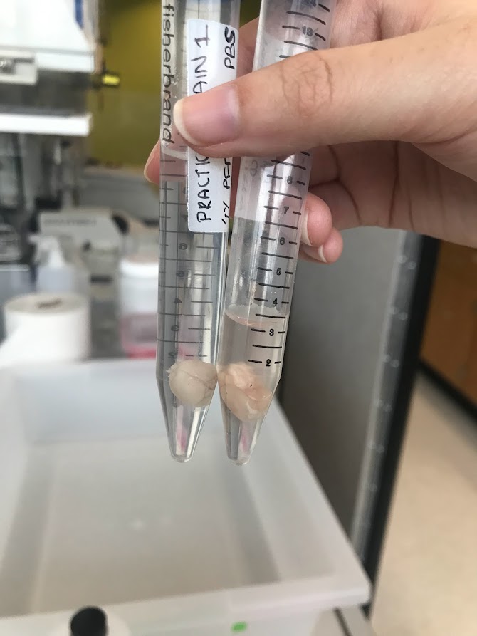
    Two mouse brains in a solution of PFA, each in its own test tube.

    After 48 hours, the brain is ready to be sliced. There’s a lot of ways to take brain slices, but we use a vibratome. First, we locate the area of the brain that we’re trying to reach. This is where a mouse atlas comes in handy. Using a small, vibrating blade, we make slices of 50 microns–or around the width of a human hair! These slices are then stained with dyes like DAPI (which makes neurons look blue)  and placed on microscope slides.

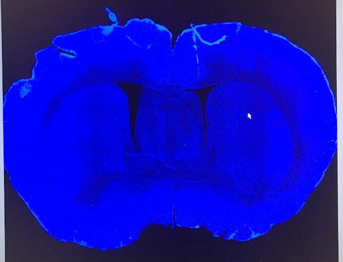
    Finally, after all this work, we can see the fruits of our labor. In our lab, we use a confocal microscope because this type of microscope can show us thin layers of cells within thick layers of tissue. Using different colors of light, we can also check how well the light sensitive proteins were able to grow in the area of the brain we’re interested in. In the picture below, you can see the first picture that I took myself from a brain I perfused and sliced. There’s no injection in this brain, so it’s as close to a “natural” brain slice that you can get. The blue color is from the DAPI dye, which indicates the location of DNA in a nucleus. So, each tiny blue dot is a neuron.
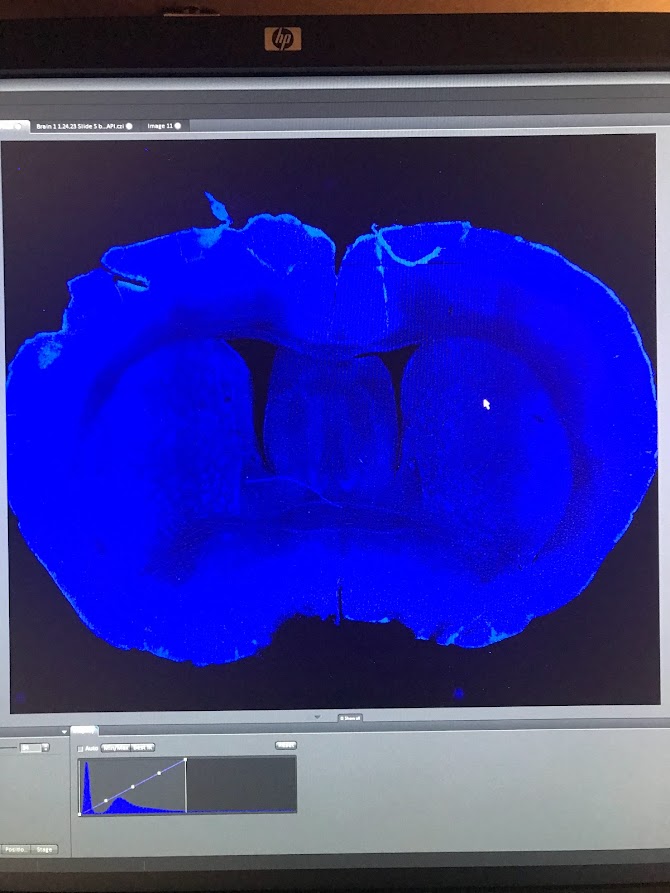
    A microscope image of the coronal slice of a mouse brain, with thousands of tiny blue dots representing individual neurons.

    It was hard to describe the first time I saw this image. Yeah, there were some nicks and tears in the tissue (my hands were shaky during the perfusion), but this was something that I had wholly produced myself. And now, in front of me, was a snapshot of all that this mouse was. 

    See that little butterfly shaped area in the middle of the brain, right above the hole in the middle? That’s the hippocampus, where every memory is scrawled in your brain through a network of neurons. What mousy memories were kept in there? Looking at this picture, I can almost imagine what it would have been like in there when the mouse was alive. The holes in between and to the side of the “wings” are ventricles, gaps in the tissue where cerebrospinal fluid carrying nutrients and hormones was kept. I can almost imagine waves of it washing over the hippocampus, keeping this great curled machine firing, shooting sparks of electricity in patterns that say I am alive. I have lived. Here is what I have seen, heard, smelled, touched, tasted, hated, loved. 

    To be honest, this work can be kind of sad. After all, all life is precious. Sometimes, it can feel almost gruesome. To perfuse a mouse, you stick a needle of fixative into its beating heart, so it evenly diffuses and preserves the entire body. The night after I read this protocol, which had a video of this process in high definition, I felt almost too nervous to sleep. When I closed my eyes, I just saw that mouse heart beating out of its chest, beating, beating, beating…

    The next morning, I admitted to my principal investigator that I wasn’t sure about trying out the perfusion that day. “Everything else, I’m okay with, but the way you can see the heart pumping freaks me out. I still want to try it, but…”

    He nodded. “You never really feel comfortable with this part of the work.”

    After a moment, he added, more seriously, “and that’s a good thing.”

    Testing on animals is a controversial subject, and I can’t claim to have the definitive opinion. But, I think my experiences give me a special point of view. In the animal labs at this institute, the animals are well cared for. Until the last day they’re with us, we treat them with respect, care, and even love (what else do you call grown adults cooing at a 2 inch long mouse washing its tiny little face with its tiny little paws?). There’s laws, regulations, committees to ensure that animals are treated ethically. They’re given pain medications after surgeries, and during procedures (surgery and perfusions) they’re put under anesthesia. Once, our lab manager stayed in the surgery room with the mouse she was operating on until night fell to make sure she was there to care for him when he woke up. 

    And yet, we have to sacrifice these animals in the end, and we have complicated feelings about it. I think that’s good and necessary. If we feel sad at the animal’s death, we treat it well when it’s alive. If we feel a bit of horror at watching its life painlessly bleed away in the dissection pan, it means we placed a high value on that life in the first place. We try to perfect our surgical skills, write better protocols, and design better experiments to minimize mistakes and the number of mice who have to undergo this process. I would much prefer that than to become cold and callous. 

    Because the truth is, we don’t have a better process to study the brain in vivo. Treatments for neurodegenerative disorders, like Parkinson’s and Huntington’s (two diseases targeting the brain area our lab studies), mental illnesses, and mental disorders lag far behind those for many other parts of the body. And the simple reason is that we often can’t get into the nervous system to study it without causing permanent damage. Until better options arise, this is the best way to perform this life-saving research.

    Whew. Things got really serious there. But working with something as big as life and death all the time makes you think about big questions. For me, ultimately this type of research is justifiable: this is the only way to validate that the research being conducted is working properly. It’s the only way to make sure that all the weird situations we put mice in weren’t done in vain. And getting to do every step of this process, going from a live mouse to a bright image on a screen–that’s something really special. Through that intricate picture, you can almost see something persisting, living on…helping us unlock the secrets of the brain. There is something to be said, here, about loving something by studying it: giving it all your attention while taking notes, appreciating the simple symmetry of the brain slice, using the gentlest touch to mount delicate tissue onto slides, admiring the natural structural variations that nature gifts every body and every brain.

    Finally, I want you to think about your daily life, and the laboratory animals that made it possible. How many medications did you take today? What about cosmetics? Vaccines? Animal testing was where many of them began. One funny thing I’ve noticed recently is that researchers who work with “animals” tend to be the most adamant about using the term “humans and non-human animals”. Other species of animals are intertwined with us in inextricable ways. And there’s really no way to really convey my gratitude to a species that can not understand it. 

    I guess this article is my attempt to try.
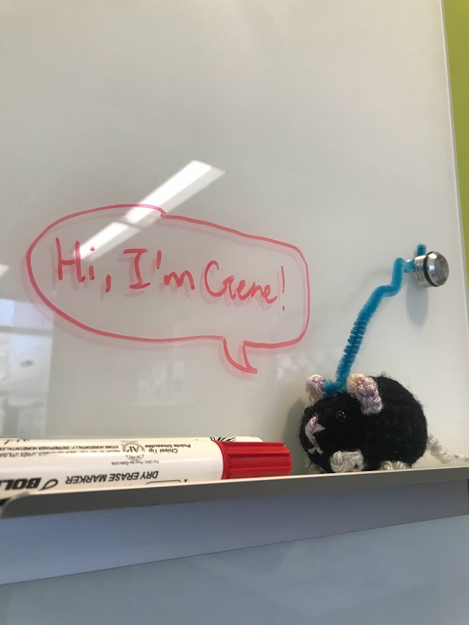
    A crochet mouse sits on the marker holder of a white board. On the white board, a text bubble reads, "Hi, I'm Gene!"

  • I Got My Brain Scanned for Science

    I Got My Brain Scanned for Science

    Yeah, I got my brain scanned for science. And you should too! (If you’re eligible, that is.)

    Basically, I got an MRI scan of my brain done recently as part of a study at the GT/GSU Center for Advanced Brain Imaging (CABI). And while I was kind of nervous about the process, I wanted to write about the experience to help other people. If you’re about to get an MRI scan for medical reasons or for a study, I hope this article can relieve any fears or answer any questions you might have!

    So, what is an MRI? MRI stands for Magnetic Resonance Imaging. An MRI machine looks like a very large tube with a donut sized hole on the end. During an MRI, you lay down in front of the donut and the technician moves you into the machine. By using magnets, the machine is able to take 3D images of your organs (in my case, my brain!). Since an MRI is so detailed and there’s no radiation risk, it is often used by doctors to detect tumors and look at brain injuries. There’s a version of an MRI called an fMRI, which shows which areas of your brain are activated. The f in fMRI stands for function, and fMRI images are able to help scientists understand how we think.

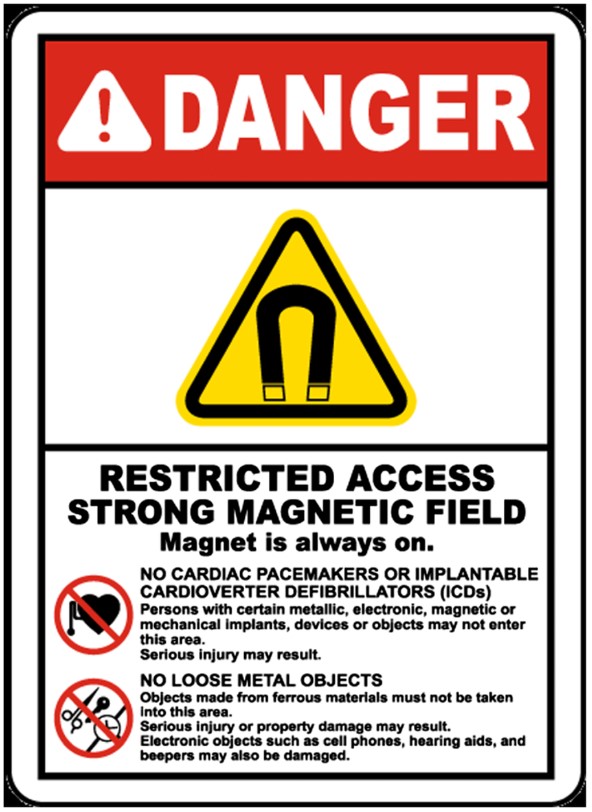
    Since an MRI uses a really powerful magnet, you can’t bring anything metal in there. So, people with pacemakers, metal stents, braces, etc. might not be able to get an MRI. And when I say a powerful magnet, I mean it. The MRI machine at CABI is behind a heavy, access locked door with two large signs looking like the one on the right:

    A danger sign with a red header saying danger. Underneath is a yellow triangle with a magnet symbol indicating a strong magnetic field.And if you have a morbid curiosity as to what would happen if you did bring in metal on or in you, check out this video: MRI vs Metal Coins (American Currency) DO NOT ATTEMPT To Replicate. Notice that just by being near the MRI, the camera goes all wonky. Some other metal things that you might not consider:

    • Some tattoo inks that include metal
    • Earrings
    • Hearing implants
    • Magnetic eyelashes
    • Those metal fasteners on pants

    If you have any of these, it doesn’t mean you’re automatically disqualified from getting an MRI, but it’s important to bring it up to the technician or radiologist.

    After I made sure I was metal free, I stepped into the MRI control room. There was a giant window where the researchers and technician would be able to see me laying in the MRI in the next room. I took off my jacket and shoes, feeling a bit jittery now. I was going to be INSIDE that giant thing.

    The research assistant made her way over to me with a Vitamin E capsule and some tape. At my confused look, she explained, “Vitamin E shows up really brightly on the MRI, so we are gonna put this on your left forehead. That way, when the scans come in, we can tell which side is the left.” Makes sense to me. But that didn’t make it feel less weird when I put it on my head.

    The technician led me into the MRI room, where I laid down on the extended platform. It felt like the kind of examination table they have at the doctor’s office, but it was more padded. That was to discourage people from moving around and messing up the scan. After I laid down, I felt the technician place down more padding around my head and feet. She also gave me earplugs and a bulb that I was supposed to squeeze if I needed anything once I was in the machine. As she did all this and wheeled me into the darkness of the machine, she told me what she was doing. This helped calm my nerves a lot, considering I couldn’t see what was going on. 

    Finally, the technician turned the machine on, and I immediately appreciated the earplugs. The beeping reminded me of an alarm clock. I heard that some people can fall asleep during an MRI, but that wasn’t me. It wasn’t earsplitting, but just loud enough that it never faded into the background.

    My earplugs were attached to an earpiece. After a burst of static, I heard the research assistant over the beeps. 

    “Okay, we’re going to do the first part of the MRI and take a picture of your brain’s structure. Do you feel okay?”

    I nodded. There was a pause. “Wait, sorry,” I said, smiling sheepishly to myself in the dark of the machine. Obviously, they couldn’t see me. “Yes, I’m all ready.”

    The actual imaging itself was boring, but I passed the time imagining the magnetic beam hitting all of my cells. After a while, I could almost feel the beam on my skin. There wasn’t actually anything, of course. Then, the fMRI was done. The beeping was a different rhythm–more pleasant, in my opinion.

    While the scan was being performed, I completed a memory task as part of the study. A screen was put in front of me while I was still in the scanner, with the image projected from a computer screen in the control room using some mirrors. Using a control pad of buttons, I was even able to manipulate what was on the screen. I didn’t even know you could do all that while in an MRI machine!

    Soon, I was able to be released from the dark void I had been in previously. The whole process took around half an hour–much shorter than I expected. Other than some numbness from staying as still as possible, I felt alright. And I was excited to see what my brain really looked like!

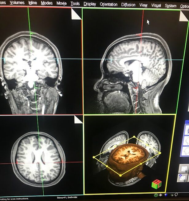
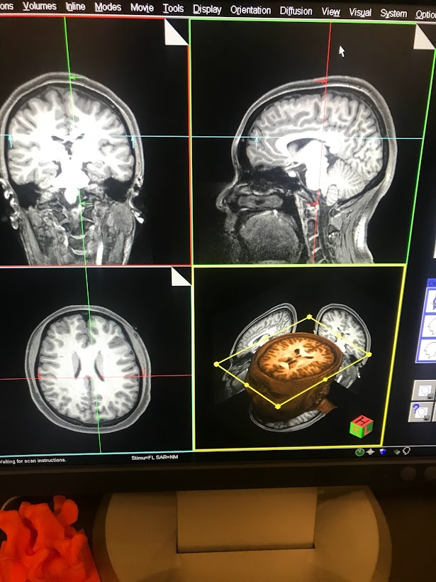
    A picture of my MRI scan from the coronal, sagittal, and horizontal views.
    This is my brain! From the top left going counterclockwise, that is the coronal view, horizontal view, and sagittal view. The bottom right shows all the slices of my brain that were taken, which you can see by moving the yellow lines.

    And there it was–all of my consciousness, my memories, my thoughts, my emotions, dreams, fears all up on that screen. It looked so complicated and fragile and beautiful. As a neuroscience major, I saw pictures of brains every day. But nothing hits quite like seeing your own brain up there in black and white. And there I was, using my brain to understand that what I saw in front of me was itself. It’s such an indescribable feeling!

    And that reason, beyond just the contributions to science, is why I recommend getting an MRI done as part of a study. After this experience, I felt myself thinking about my own brain with more affection. This squishy, bumpy organ is me, and I should treat it with the study and love that it deserves.


    References

    https://www.researchgate.net/figure/Magnetic-field-warning-sign_fig1_345758192

  • Neurotransmitter Notes: Serotonin + Oxytocin

    Neurotransmitter Notes: Serotonin + Oxytocin

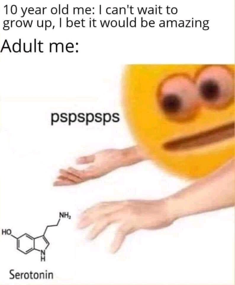
    10 year old me: I can't wait to grow up, I bet it would be amazing. Adult me: a picture of a cartoon face making the pspsps sound at a chemical diagram of serotonin the way one would call a cat.


    Take two of our Neurotransmitter Notes! If you are in the midst of finals season like me, maybe you want something to cheer you up. I might not be able to make your neurons fire off serotonin or oxytocin, but maybe learning about them will help you produce your own! Without further ado, let’s set the tone with serotonin…


    “Am I going crazy

    Consuming me lately

    And I’m well

    I’m over it

    Not where I’ve been

    It’s where I’m going

    Looking for my vibe

    Wanna bathe myself in serotonin”


    Serotonin- Courtney Govan


    After dopamine, serotonin might just be the second most well known neurotransmitter. And like dopamine, it has effects beyond making you happier: it could be better described as a mood regulator that can also affect your sleep, digestion, and wound healing (Serotonin, n.d.).


    But before you buy those serotonin gummies off the internet, be careful! Too much serotonin can lead to serotonin syndrome, This alliterative ailment can cause vomiting, headache, rapid heart rate, and muscle twitches (Dunkin, n.d.). In this situation, you really can have too much of a good thing. But what about the opposite?


    A lot of people being treated for depression know about serotonin in the context of “the thing I need more of”. This perception is described in girl in red’s Serotonin:


    “I’m running low on serotonin

    Chemical imbalance got me twisting things

    Stabilize with medicine

    But there’s no depth to these feelings

    Dig deep, can’t hide

    From the corners of my mind

    I’m terrified of what’s inside”


    Serotonin- girl in red


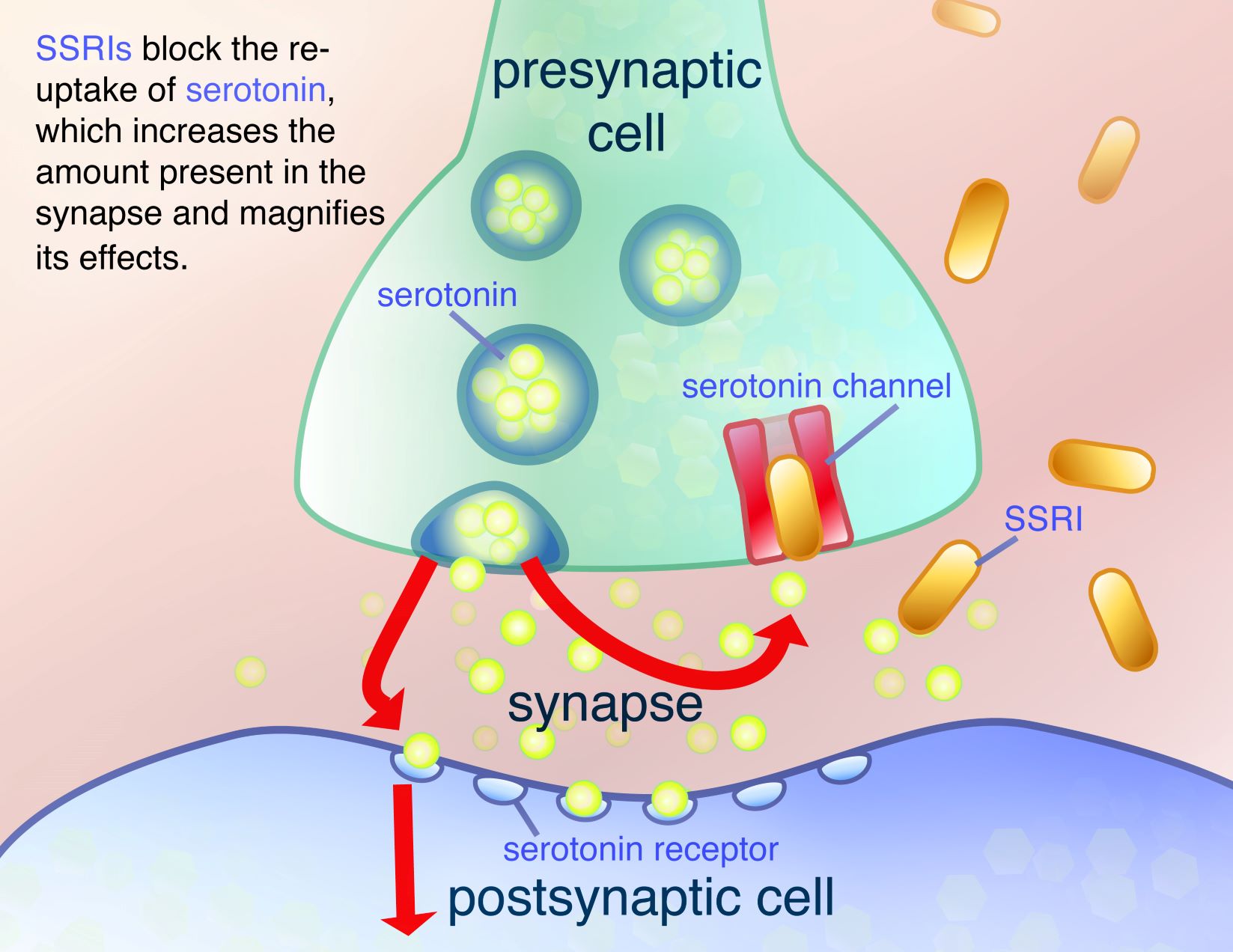
    Indeed, depression is linked to lower activity of neurons that use serotonin. One of the most common antidepressants prescribed nowadays are the selective-serotonin reuptake inhibitors (SSRIs). The medicine girl in red describes is likely an SSRI, which works by preventing serotonin from being taken up by neurons after it is released into a synapse (the junction between neurons where neurotransmitters transfer a signal from one neuron to the next). This increases synaptic serotonin levels. Then, like if you were to increase the number of balls in a pinball machine, the serotonin is more likely to bind to a receptor on the other side, activating the next neuron. Boom–more activity!

    an image showing the process by which ssris prevent serotonin from leaving the synapse.
    A diagram showing the function of SSRIs (SSRIs, 2010)


    And to round out our discussion of the three “happy chemicals” people know about, let’s talk about oxytocin! Now, oxytocin is technically a hormone and not a neurotransmitter (do you remember the difference from my last article?).  Compared to dopamine and serotonin, oxytocin is perhaps the one most related to gettin’ hot and heavy in the public eye. Just take a look at these lyrics from Billie Eilish:


    “Can’t take it back once it’s been set in motion

    You know I need you for the oxytocin

    If you find it hard to swallow

    I can loosen up your collar

    ‘Cause as long as you’re still breathing

    Don’t you even think of leaving”


    Oxytocin- Billie Eilish


    Here, she really shows the burst of excitement that can stem from oxytocin. Oxytocin makes us feel more connected to the people around us. However, it is not just released among lovers–it’s actually a key player in childbirth and nursing (Watson, 2021). It is also released during exercise, listening to music, and any type of physical contact. How touching!


    That’s all I have for you all today. Take some personal time to relax and hug a friend (or partner)! We’ll get through this period together.


    Check out this playlist with the tunes mentioned in this article, as well as some other neuroscience related songs! (Note: please be aware that some songs in this playlist may have explicit lyrics)


    References

    Dunkin, M. A. (n.d.). Serotonin syndrome. WebMD. Retrieved April 27, 2022, from https://www.webmd.com/depression/guide/serotonin-syndrome-causes-symptoms-treatments

    Serotonin: What is it, function & levels. (n.d.). Cleveland Clinic. Retrieved April 27, 2022, from https://my.clevelandclinic.org/health/articles/22572-serotonin

    Skabreus. (2021, June 12). I can’t wait to grow up [Reddit Post]. R/Memes. www.reddit.com/r/memes/comments/ny9ds6/i_cant_wait_to_grow_up/

    SSRIs. (2010, June 30). HOPES Huntington’s Disease Information. https://hopes.stanford.edu/ssris/

    Watson, S. (2021, July 20). Oxytocin: The love hormone. Harvard Health. https://www.health.harvard.edu/mind-and-mood/oxytocin-the-love-hormone


  • Neurotransmitter Notes: Dopamine

    Neurotransmitter Notes: Dopamine

    “I’m just a chemical in your head

    I’m just that Dopamine

    So come on line me up

    Come on line me up”

    – “Dopamine”, Third Eye Blind

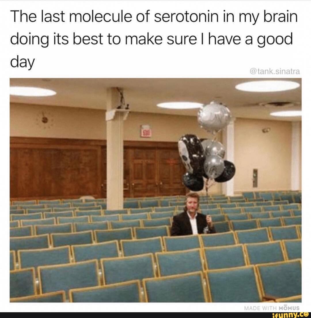
    As a neuroscience major, it makes me extremely happy when words about the brain and nervous system enter the popular vocabulary. And recently, I’ve noticed people have been using neurotransmitters to talk about their feelings. If someone wants to say something brings them joy, they’ll say, “That’s my dopamine!”. Or, if someone feels unhappy, they might share a meme about how they want more serotonin (like the meme below!)

    There is a picture of a man sitting alone in a lecture hall holding a bunch of balloons. The caption reads the last molecule of serotonin in my brain doing its best to make sure I have a good day.

    But to me, the biggest indicator of whether something has entered the popular consciousness is if there’s music about it. So, through the article series “Neurotransmitter Notes”, I want to chat about songs that talk about the 8 major neurotransmitters: epinephrine, norepinephrine, dopamine, serotonin, GABA, acetylcholine, glutamate, and endorphins. Today, we’ll tackle the most famous neurotransmitter of them all: dopamine.

    Neurotransmitters

    Hold on, what’s a neurotransmitter? The Encyclopedia Brittanica defines a neurotransmitter as “any of a group of chemical agents released by neurons (nerve cells) to stimulate neighbouring neurons or muscle or gland cells, thus allowing impulses to be passed from one cell to the next throughout the nervous system” (Neurotransmitter | Definition, Signaling, & Types | Britannica, n.d.). So, neurotransmitters (NTs) are chemical messengers that neurons use to pass signals onto one another. What differentiates NTs from one another are that NTs will only be sent and received by the neurons that have the protein receptors that fit with it. Neurons are classified by the type of neurotransmitter it can “understand”: so, you’ll have dopaminergic neurons (which send and receive signals about dopamine). Those dopaminergic neurons, when binding to dopamine, will cause the effects that are associated with dopamine.

    Dopamine’s Effects

    Like I mentioned earlier, dopamine is known as the “happy” chemical. But it’s not just released when you feel good: it plays many, many other roles. One role is controlling decision making. For instance, higher levels of it are related to whether someone chooses to do a more difficult, rewarding task over a simpler, less rewarding one (Dopamine Affects How Brain Decides Whether a Goal Is Worth the Effort, 2020). These facts, I’m sure, were handy when writing the lyrics to this dance jam:


    “You make all the dopa-dopamine in my bloodstream

    The way you touch me, I’m loving the feeling

    Of dopa-dopamine in my bloodstream

    Let it control me, I’m loving the feeling”

    – “Dopamine”, Purple Disco Machine ft. Eyelar


    The songwriters have demonstrated here the happy feeling associated with dopamine, as well as its importance in controlling behavior. However, there’s also an error: neurotransmitters don’t travel in the bloodstream. Dopamine happens to be unique because it has an alternate form that does travel in the bloodstream, but that form doesn’t have the same effects as what the song’s describing. It’s likely that they mixed up neurotransmitters with hormones, which do travel in the bloodstream.

    Dopamine Doesn’t Just Move You, It Motivates You

    Not only can dopamine make you feel good and affect your decision making, it can impact your motivation. This motivation can be in the form of how well you can focus and retain information for a task (executive function). Executive dysfunction occurs in people with ADHD, who also often have lower levels of dopamine in their brain (Logue & Gould, 2014). 

    Motivation also plays a role in addiction. The ventral tegmental area (VTA) is a section of neurons in the brain that play a major role in people with addictions, and these neurons are dopaminergic. In the song “Dopamine” by Bliss n Eso, the singer talks about his reliance on alcohol to help him through times of stress. This chorus describes how it felt for him to use it:


    “When there’s no way out

    When I don’t believe

    When I’m coming down

    You’re my dopamine”

    – “Dopamine”, Bliss n Eso


    However, over the course of the song, he cites his connections to his wife and child as motivation to become a better person. And by the end, this same chorus has taken on a new meaning. By the way, long-term intense romantic love still makes your VTA light up with dopamine, years after the honeymoon period has ended (Acevedo et al., 2012).

    Dope-amine or Nope-amine?

    I suppose we can take from this a lesson: dopamine is not just a feel-good chemical– it’s crucial for the proper functioning of your brain. Don’t try to chase that short-term high: if you take care of yourself and make meaningful connections to other people, dopamine (and life satisfaction) will come to you.

    Check out this playlist with the tunes mentioned in this article, as well as some other neuroscience related songs! (Note: please be aware that some songs in this playlist may have explicit lyrics.) I’ll be back soon with some more Neurotransmitter Notes!


    References

    Acevedo, B. P., Aron, A., Fisher, H. E., & Brown, L. L. (2012). Neural correlates of long-term intense romantic love. Social Cognitive and Affective Neuroscience, 7(2), 145–159. https://doi.org/10.1093/scan/nsq092

    Dopamine affects how brain decides whether a goal is worth the effort. (2020, March 30). National Institutes of Health (NIH). https://www.nih.gov/news-events/nih-research-matters/dopamine-affects-how-brain-decides-whether-goal-worth-effort

    Logue, S. F., & Gould, T. J. (2014). The neural and genetic basis of executive function: Attention, cognitive flexibility, and response inhibition. Pharmacology, Biochemistry, and Behavior, 0, 45–54. https://doi.org/10.1016/j.pbb.2013.08.007

    Neurotransmitter | definition, signaling, & types | britannica. (n.d.). Retrieved February 19, 2022, from https://www.britannica.com/science/neurotransmitter

  • My Experience as a STEM Program Planning Assistant

    My Experience as a STEM Program Planning Assistant

    For people who are interested in STEM, there are a variety of jobs and experiences you can have. For me, an undergraduate student at Georgia Tech, I think I have an interesting job that goes along with my coursework and other activities. Since it’s a pretty unique position, I thought I’d share my experiences and reflections.

    First, I mentioned in the title that I work as a STEM program’s planning assistant. Specifically, I work for the Explore Living Learning Community (LLC) at Georgia Tech, a program for first years where students live together, take similar classes, and get involved in activities hosted by the LLC. These activities include educational events and social opportunities, which are planned and hosted by student assistants who are Tech students too! This LLC focuses on pre-health and research, and most of our students are STEM majors.

    I’ve had this position for two semesters now, and I’ve found it a great opportunity to combine my love of STEM with science communication. Paying close attention to what students are interested in then creating engaging and useful events requires STEM knowledge, familiarity with STEM students, and plenty of soft skills that aren’t typically trained as part of a science education. But, they have wide applicability. For instance, you can imagine any of these skills coming in handy whether you’re a scientist applying for a grant or an engineer trying to sell your new invention:

    • Event planning
    • Communication skills with clients, coworkers, and superiors
    • Marketing knowhow

    In addition to these soft skills, I’ve been able to take a peek into what the next generation of scientists, engineers, and programmers tick. They want to try new things that may not be directly related to their program of study. They aspire to make professional connections in their field to gain an edge. And, they’re eager to participate in events that will foster their curiosity and intellectual growth. To help them achieve this, our program has done a plethora of activities from paint nights, research networking events, and even a popular Halloween trivia night that I put on.

    What touched me most about my work so far is the possibility for students to feel inspired by STEM again. Coming on the third year of the pandemic, a lot of my fellow students feel quite burnt out from school, work, coronavirus, etc. In these circumstances, it’s hard for someone to stay motivated to the demanding area of study that they decided to pursue. But by connecting their areas of study with community, career, and fun, we can bring back in small part that little spark that made us interested in all this in the first place.

    One of my first major activities this fall was leading a trip to the Atlanta Zoo. After getting there, most of the students split up into groups to explore the park. I was left with a handful of guys who didn’t seem to have a group to go with. As expected, things were very quiet and shy, and I was a little scared at first that they might not enjoy the experience. After all, how can you stay stony faced when you’re looking straight at an elephant giving itself a dust bath?

    But after about an hour, my fears were quelled. They started smiling, laughing, and telling jokes. By lunch, we were thick as thieves. It’s hard to tell what caused this shift. The nice long walk? The cute cool looking animals? The real-world not video call social interaction?

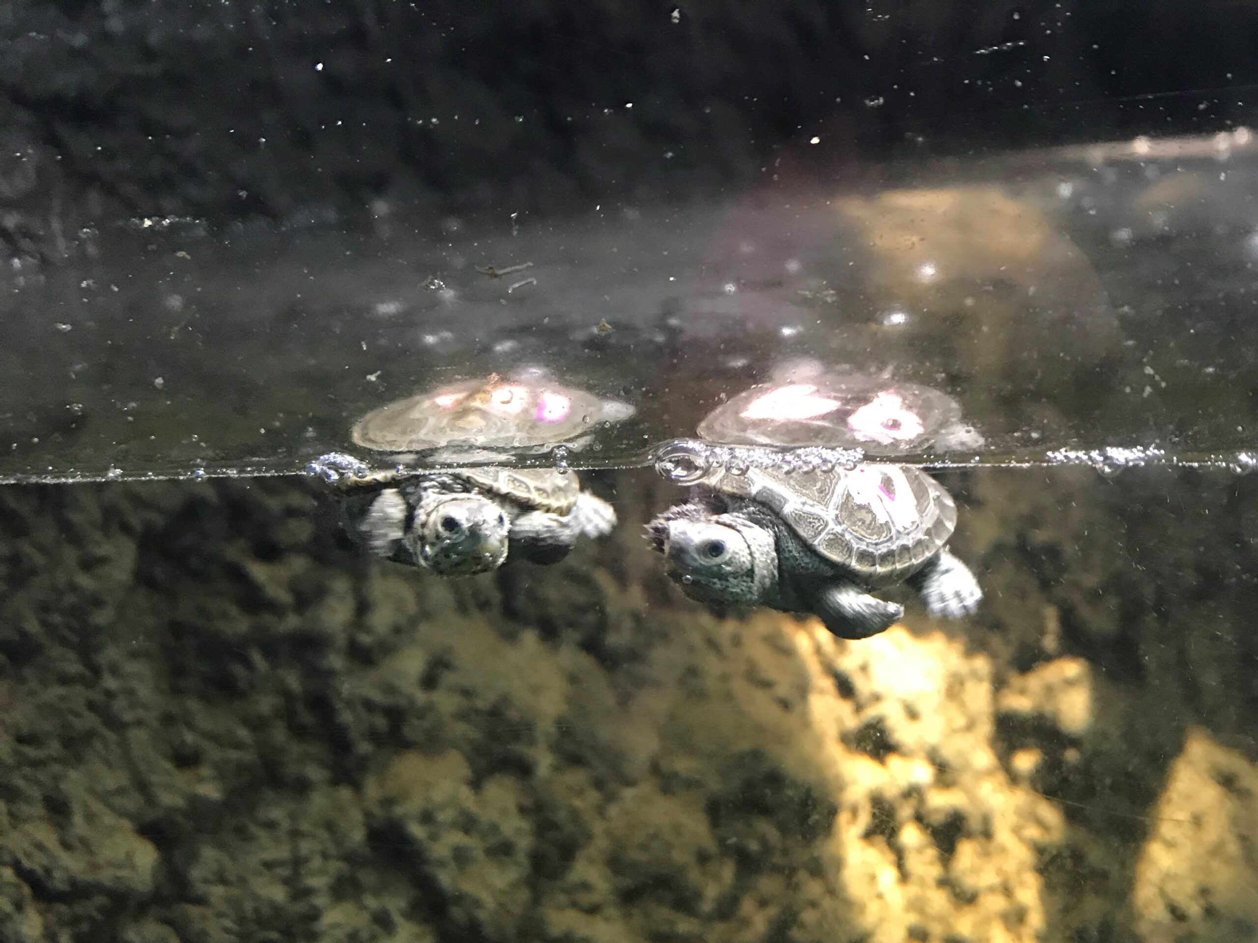
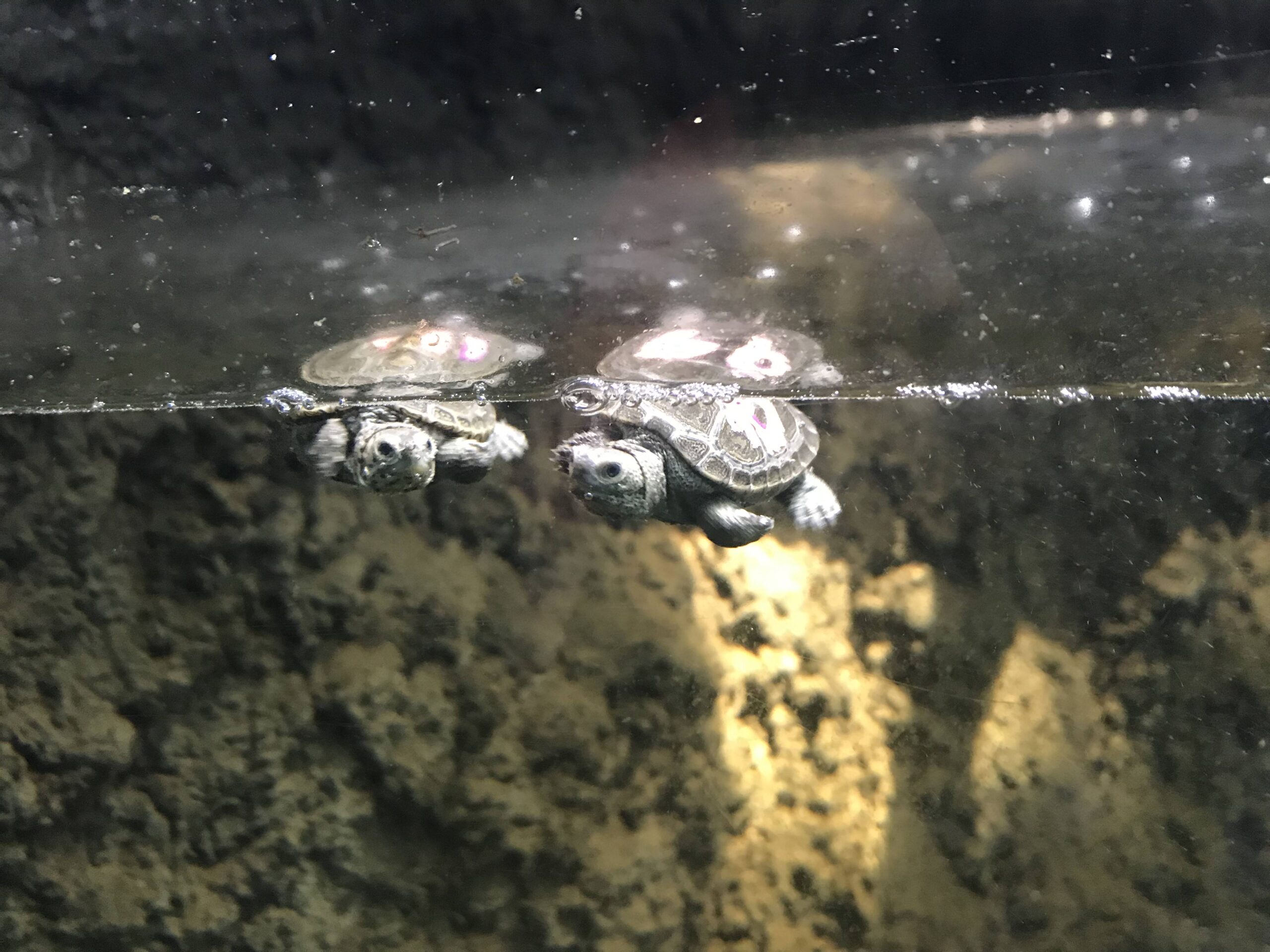
    In the reptile exhibit, seeing these previously deadpan students coo and aww over baby turtles, I mused to myself that we’d rediscovered something. One student remarked that after all those exams, seeing them made him feel like everything was okay again. You could see that spark in their eyes, the one that we all had when we were kids and we found something wonderful. After all, it was first our curiosity and sense of wonder that led us to STEM to seek out answers. And can’t we all use a reminder once in a while?

  • ‘Vampire Facials’: Viable or Just Bloody Gross?

    ‘Vampire Facials’: Viable or Just Bloody Gross?

    To pursue beauty, people will use makeup, creams, and even plastic surgery. But, would you spread your own blood on your face?

    Wait, Your Own Blood?

    Earlier this year, these treatments, known as ‘blood facials’ or ‘vampire facials’ became well known due to celebrities such as Kim Kardashian posting about their rejuvenation effect on social media. Quickly, this sparked a debate over whether these treatments were handy or horrific. And who knows if they even work?

    Well, Do They Work?

    These vampire facials are actually known clinically as protein-rich plasma (PRP) treatments. First, your blood is drawn, then spun through a centrifuge. The high force makes the blood separate into three layers: red blood cells at the bottom, liquid plasma at the top, and a cloudy layer called the “serum portion” in the middle that contains the protein-rich plasma. After processing the PRP, it is ready for treatment. Treatment can include injecting it under the skin the way one would inject fillers, or it can be applied like a face mask. If the PRP is applied, it can be combined with a procedure like microneedling or microabrasion, which open up little holes and cracks in the skin to help the plasma absorb into the face. The dermatologists who perform them promise effects like reversal of hair loss, increased collagen production, and glowing skin. 

    Just a Selling Point?

    While there aren’t many clinical studies on these effects yet, PRP treatments have actually been used for healing joint and muscle injuries. They can speed up cell regeneration for a variety of sports injuries and even alleviate chronic tendonitis pain. The process is quite similar to how vampire facials work. The platelet-rich plasma is just injected or applied onto the skin of the area that needs to grow back.

    Tell Me More!

    For both the cosmetic and medical treatments, the plasma is full of platelets. In the blood, they help it clot when you get a cut. They contain alpha granules that have growth factors in them. These growth factors act as chemical messengers to promote cell division and growth. This also includes producing more proteins such as collagen, which provides structure to the body. So, they can help a torn ACL reform or cause cheek cells to multiply and plump up a face. Since PRP is minimally invasive and has few risks, it can be a good option for patients who have a variety of treatment goals.

    If You’re Still Wrapping Your Head Around It All, You’re Not Alone!

    Many people can get put off by this bloody procedure. On the surface, it sounds like yet another one of those beauty gimmicks that seems to exist only to trick the wealthy out of their money. But astonishingly, protein-rich plasma treatments have a basis in regenerative medicine. In reality, the worlds of beauty, science, and medicine can be very intertwined. When these disciplines learn from one another, interesting and helpful things can happen. What other examples of this other than vampire facials can you think of?

    In any case, happy (belated) Halloween. Remember, your blood is very, very precious.


    Citations

    Abelman, D. (2021, August 5). What Exactly Is a Vampire Facial? . Allure. Retrieved November 1, 2021, from https://www.allure.com/story/vampire-facial-prp-treatment-guide.

    Platelet-rich plasma (PRP) injection: How it works: HSS. Hospital for Special Surgery. (n.d.). Retrieved November 1, 2021, from https://www.hss.edu/condition-list_prp-injections.asp.

    Setayesh, K., Villarreal, A., Gottschalk, A., Tokish, J. M., & Choate, W. S. (2018, December). Treatment of muscle injuries with platelet-rich plasma: A review of the literature. Current reviews in musculoskeletal medicine. Retrieved November 1, 2021, from https://www.ncbi.nlm.nih.gov/pmc/articles/PMC6220013/.Â